Assessing the application of tissue microarray technology to kidney research
- PMID: 20086233
- PMCID: PMC2857813
- DOI: 10.1369/jhc.2009.954966
Assessing the application of tissue microarray technology to kidney research
Abstract
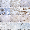
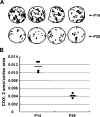
Tissue microarray (TMA) is a new high-throughput method that enables simultaneous analysis of the profiles of protein expression in multiple tissue samples. TMA technology has not previously been adapted for physiological and pathophysiological studies of rodent kidneys. We have evaluated the validity and reliability of using TMA to assess protein expression in mouse and rat kidneys. A representative TMA block that we have produced included: (1) mouse and rat kidney cortex, outer medulla, and inner medulla fixed with different fixatives; (2) rat kidneys at different stages of development fixed with different fixatives; (3) mouse and rat kidneys with different physiological or pathophysiological treatments; and (4) built-in controls. As examples of the utility, immunostaining for cyclooxygenase-2, renin, Tamm Horsfall protein, aquaporin-2, connective tissue growth factor, and synaptopodin was carried out with kidney TMA slides. Quantitative analysis of cyclooxygense-2 expression in kidneys confirms that individual cores provide meaningful representations comparable to whole-kidney sections. These studies show that kidney TMA technique is a promising and useful tool for investigating the expression profiles of proteins of interest in rodent kidneys under different physiological and pathophysiological conditions.
Figures


Similar articles
-
Defective expression of Tamm-Horsfall protein/uromodulin in COX-2-deficient mice increases their susceptibility to urinary tract infections.Am J Physiol Renal Physiol. 2005 Jul;289(1):F49-60. doi: 10.1152/ajprenal.00134.2004. Epub 2005 Mar 1. Am J Physiol Renal Physiol. 2005. PMID: 15741608
-
Ultrastructural localization of Tamm-Horsfall glycoprotein (THP) in rat kidney as revealed by protein A-gold immunocytochemistry.Histochemistry. 1985;83(6):531-8. doi: 10.1007/BF00492456. Histochemistry. 1985. PMID: 3910623
-
Renal effects of Tamm-Horsfall protein (uromodulin) deficiency in mice.Am J Physiol Renal Physiol. 2005 Mar;288(3):F559-67. doi: 10.1152/ajprenal.00143.2004. Epub 2004 Nov 2. Am J Physiol Renal Physiol. 2005. PMID: 15522986
-
Validation of 2-mm tissue microarray technology in gastric cancer. Agreement of 2-mm TMAs and full sections for Glut-1 and Hif-1 alpha.Anticancer Res. 2014 Jul;34(7):3313-20. Anticancer Res. 2014. PMID: 24982335
-
Making a Tissue Microarray.Methods Mol Biol. 2019;1897:313-323. doi: 10.1007/978-1-4939-8935-5_27. Methods Mol Biol. 2019. PMID: 30539455 Review.
Cited by
-
Resistance to hypertension mediated by intercalated cells of the collecting duct.JCI Insight. 2017 Apr 6;2(7):e92720. doi: 10.1172/jci.insight.92720. JCI Insight. 2017. PMID: 28405625 Free PMC article.
-
New isoform-specific monoclonal antibodies reveal different sub-cellular localisations for talin1 and talin2.Eur J Cell Biol. 2012 Mar;91(3):180-91. doi: 10.1016/j.ejcb.2011.12.003. Epub 2012 Feb 3. Eur J Cell Biol. 2012. PMID: 22306379 Free PMC article.
References
-
- Chen XM, Qi W, Pollock CA (2009) CTGF and chronic kidney fibrosis. Front Biosci 1:132–141 - PubMed
-
- Durvasula RV, Shankland SJ (2006) Podocyte injury and targeting therapy: an update. Curr Opin Nephrol Hypertens 15:1–7 - PubMed
-
- Harris RC, Zhang MZ, Cheng HF (2004) Cyclooxygenase-2 and the renal renin-angiotensin system. Acta Physiol Scand 181:543–547 - PubMed
MeSH terms
Substances
LinkOut - more resources
Full Text Sources
Research Materials

