. 2010 Jan 19;3(105):ra4.
doi: 10.1126/scisignal.2000567.

Distinct signal codes generate dendritic cell functional plasticity

Affiliations

Distinct signal codes generate dendritic cell functional plasticity

Kazuhiko Arima et al. Sci Signal. .

Abstract

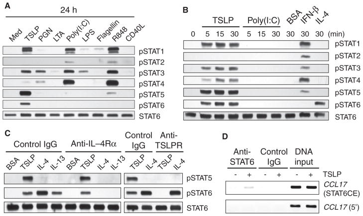
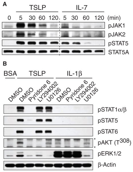
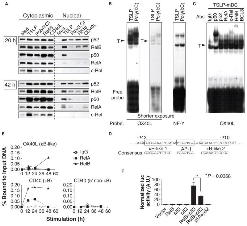
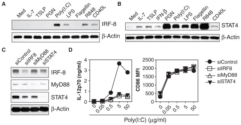
Our adaptive immune system induces distinct responses to different pathogens because of the functional plasticity of dendritic cells (DCs); however, how DCs program unique responses remains unclear. Here, we found that the cytokine thymic stromal lymphopoietin (TSLP) potently transduced a unique T helper type 2 (T(H)2)-inducing compound signal in DCs. Whereas activation of nuclear factor kappaB (predominantly p50) drove DCs to produce OX40L to induce T(H)2 differentiation, the activation of signal transducer and activator of transcription 6 (STAT6) triggered DCs to secrete chemokines necessary for the recruitment of T(H)2 cells. In addition, TSLP signaling limited the activation of STAT4 and interferon regulatory factor 8 (IRF-8), which are essential factors for the production of the T(H)1-polarizing cytokine interleukin-12 (IL-12). By contrast, Toll-like receptor ligands and CD40 ligand did not activate STAT6 in myeloid DCs, but instead increased the abundance of STAT4 and IRF-8 to induce T(H)1 responses through the production of IL-12. Therefore, we propose that the functional plasticity of DCs relies on elaborate signal codes that are generated by different stimuli.


Figures

Fig. 1
Fig. 1
TSLP directly activates STAT6 in mDCs. (A) Western blotting analysis was performed to compare the activation of different STAT proteins in mDCs cultured for 24 hours with medium alone (Med), TSLP (25 ng/ml), PGN, LTA, poly(I:C), LPS, flagellin, R848, or CD40L. (B) Western blotting analysis was performed to compare the early time course of activation of STAT proteins in mDCs stimulated with TSLP (25 ng/ml), poly(I:C), vehicle control (BSA), IFN-β (25 ng/ml), or IL-4 (25 ng/ml). (C) Effect of neutralizing antibodies against IL-4Rα or TSLPR on the activation of the indicated STAT proteins in mDCs by TSLP, IL-4, and IL-13. Cells were preincubated with the indicated neutralizing or isotype-matched control antibodies (10 μg/ml) for 30 min at 37°C and then stimulated with 10 ng/ml of TSLP, IL-4, or IL-13 or with vehicle control (BSA) for 10 min at 37°C. (A to C) Western blots were incubated with antibodies specific for the indicated phosphorylated STAT proteins or with an antibody against STAT6 to confirm equal loading of all lanes. Data are representative of three independent experiments. (D) ChIP assays demonstrating the enrichment of STAT6 at the region around the STAT6 consensus elements (STAT6CE) of the promoter of CCL17 in response to TSLP. A region ~6000 bp upstream of the initiator codon of the gene was used as negative control. Data are displayed as inverted images for easier visibility and are representative of three independent experiments. IgG, immunoglobulin G.
Fig. 2
Fig. 2
Strong activation of JAK mediates sustained and broad TSLP-dependent signaling. (A) Western blotting analysis was performed to compare the kinetics of TSLP- and IL-7–mediated phosphorylation of JAK1, JAK2, and STAT5 in mDCs. Western blots were incubated with specific antibodies against the indicated phosphorylated proteins or with an antibody against STAT5A to confirm equal loading of all lanes. (B) Western blotting analysis was performed to demonstrate the contributions of JAK, PI3K, and ERK to TSLP-mediated signals. mDCs were pretreated with the pan-JAK inhibitor pyridone 6, the PI3K inhibitor LY294002, the MEK (ERK) inhibitor U0126, or DMSO (0.1%) for 30 min at 37°C and then were stimulated with 10 ng/ml of TSLP or IL-1β or with BSA for 10 min at 37°C. (*) Nonspecific bands. (A and B) The data shown are representative of three independent experiments.
Fig. 3
Fig. 3
TSLP activates NF-κB–dependent pathways that lead to the increased abundance of OX40L at the cell surface of mDCs. (A) Western blotting analysis was performed to demonstrate the cytoplasmic and nuclear localization of NF-κB molecules in mDCs 20 and 42 hours after culture with medium alone (Med), TSLP, poly(I:C), R848, or CD40L. (B) EMSA showing the binding of nuclear proteins to probes for the κB-like sequence of the promoter of OX40L and the binding sequence of the “housekeeping” transcription factor NF-Y in mDCs cultured for 60 hours with TSLP or poly(I:C). For the experiment with the OX40L probe, an autoradiograph of the same gel after a shorter exposure time is added to show the band separation. T, TSLP-predominant bands. (C) Super-shift assays were performed with antibodies against the indicated NF-κB molecules and BCL3 to demonstrate the composition of the nuclear proteins bound to the κB-like sequence probe of OX40L from mDCs cultured for 60 hours with TSLP. (D) Nucleotide sequence of the region surrounding the potential NF-κB binding sites within the 5′-flanking region of OX40L. The nucleotide positions indicate the relative distance from the initiator codon. The sequences of the consensus κB site and the activating protein 1 (AP-1) site are depicted. (E) ChIP assays demonstrating the kinetics of the recruitment of RelA and RelB to the promoters of OX40L and CD40 in TSLP-treated mDCs. Data are expressed as the percentage binding of either protein to the input DNA amount by quantifying the intensities of PCR-amplified bands. (A to C, and E) Data are representative of three independent experiments. (F) NF-κB–dependent activation of the OX40L promoter in HEK 293T cells was determined by luciferase reporter assay. Error bars represent the SEM from three independent experiments.
Fig. 4
Fig. 4
TSLP does not stimulate the production of IRF-8 or STAT4, essential factors for the production of IL-12 by mDCs. (A) Western blotting analysis was performed to compare the differential regulation of the abundance of IRF-8 in mDCs that were activated for 24 hours by the indicated stimuli, as described in Fig. 1A with the inclusion of IL-7 (25 ng/ml) and the exclusion of LTA. (B) Western blotting analysis was performed to compare the abundance of STAT4 in mDCs after 24 hours of activation by the indicated stimuli, as described in (A) with the inclusion of IFN-β (25 ng/ml).(C) Western blotting analysis was performed to show the efficiency of knockdown of the indicated proteins in poly(I:C) (5 μg/ml)-stimulated mDCs transfected with siRNAs specific for IRF-8, MyD88, or STAT4 or with a nontargeting control siRNA (siControl). (A to C) Western blots were incubated with antibody against β-actin to demonstrate equivalent loading of all lanes. (D) siRNA-transfected mDCs were stimulated with poly(I:C) for 24 hours and the concentration of IL-12p70 in the culture supernatants and the mean fluorescent intensity (MFI) of cell-surface CD86 are depicted. Data are representative of three (A and B) or five (C and D) independent experiments.

References

    1. Banchereau J, Briere F, Caux C, Davoust J, Lebecque S, Liu YJ, Pulendran B, Palucka K. Immunobiology of dendritic cells. Annu Rev Immunol. 2000;18:767–811. - PubMed
    1. Macatonia SE, Hosken NA, Litton M, Vieira P, Hsieh CS, Culpepper JA, Wysocka M, Trinchieri G, Murphy KM, O’Garra A. Dendritic cells produce IL-12 and direct the development of Th1 cells from naive CD4+ T cells. J Immunol. 1995;154:5071–5079. - PubMed
    1. Cella M, Scheidegger D, Palmer-Lehmann K, Lane P, Lanzavecchia A, Alber G. Ligation of CD40 on dendritic cells triggers production of high levels of interleukin-12 and enhances T cell stimulatory capacity: T-T help via APC activation. J Exp Med. 1996;184:747–752. - PMC - PubMed
    1. Kaliński P, Hilkens CM, Wierenga EA, Kapsenberg ML. T-cell priming by type-1 and type-2 polarized dendritic cells: The concept of a third signal. Immunol Today. 1999;20:561–567. - PubMed
    1. Moser M, Murphy KM. Dendritic cell regulation of TH1-TH2 development. Nat Immunol. 2000;1:199–205. - PubMed