Clinical Trial
. 2010 Feb;66(2):323-32; discussion 332.
doi: 10.1227/01.NEU.0000360379.95800.2F.

Transcranial magnetic resonance imaging- guided focused ultrasound surgery of brain tumors: initial findings in 3 patients

Affiliations
Clinical Trial

Transcranial magnetic resonance imaging- guided focused ultrasound surgery of brain tumors: initial findings in 3 patients

Nathan McDannold et al. Neurosurgery. 2010 Feb.

Abstract

Objective: This work evaluated the clinical feasibility of transcranial magnetic resonance imaging-guided focused ultrasound surgery.

Methods: Transcranial magnetic resonance imaging-guided focused ultrasound surgery offers a potential noninvasive alternative to surgical resection. The method combines a hemispherical phased-array transducer and patient-specific treatment planning based on acoustic models with feedback control based on magnetic resonance temperature imaging to overcome the effects of the cranium and allow for controlled and precise thermal ablation in the brain. In initial trials in 3 glioblastoma patients, multiple focused ultrasound exposures were applied up to the maximum acoustic power available. Offline analysis of the magnetic resonance temperature images evaluated the temperature changes at the focus and brain surface.

Results: We found that it was possible to focus an ultrasound beam transcranially into the brain and to visualize the heating with magnetic resonance temperature imaging. Although we were limited by the device power available at the time and thus seemed to not achieve thermal coagulation, extrapolation of the temperature measurements at the focus and on the brain surface suggests that thermal ablation will be possible with this device without overheating the brain surface, with some possible limitation on the treatment envelope.

Conclusion: Although significant hurdles remain, these findings are a major step forward in producing a completely noninvasive alternative to surgical resection for brain disorders.


Figures

Figure 1
Figure 1
Diagram of the TcMRgFUS device for noninvasive brain tumor ablation.
Figure 2
Figure 2
Screenshots from TcMRgFUS treatment planning workstation. (A) Coronal T2-weighted images of the patient in the TcMRgFUS device. The target of the current sonication is indicated by the blue rectangle. The water filling the space between the patient’s shaved head and the transducer can be seen. (B) Pre-treatment CT scan data of the skull is registered the intra-treatment MRI scans. The skull is automatically segmented from the CT scan and displayed on top of the MRI images used for treatment planning as a green region. Any registration errors can be seen on these images and corrected by the user using a graphical tool. MR tracking coils integrated into the transducer are used to register the TcMRgFUS system coordinates with the imaging coordinates. Acoustic models taking into account the patient-specific skull geometry and density are used to correct for aberrations to the ultrasound beam. (C) The beam paths for each phased array element are superimposed on the images, allowing the user to verify that no beams pass through undesired structures. (D)-(E). Pre-treatment contrast-enhanced T1-weighted images, which can be useful to define tumor margins, acquired the day before treatment can also be registered to the intra-treatment images. Axial and sagittal images are also acquired, allowing for treatment planning in three dimensions. (F). Sagittal T2-weighted image.
Figure 3
Figure 3
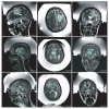
T2-weighted FSE images of the patients within the TcMRgFUS device. The outlines approximately delineate the boundaries of the targeted tumors. The space between the patients’ shaved heads and the hemispherical transducer was filled with chilled water that was continuously degassed and circulated before and between sonications. (Top: patient #1, middle: patient #2, right: patient #3; left: sagittal images, center: axial images, right: coronal images)
Figure 4
Figure 4
(A)-(D) Focal heating during TcMRgFUS in the three patients. (A)-(B): Sagittal (A) and axial (B) examples of MRTI acquired at peak temperature rise during two 20 s sonications (acoustic power: 800 W) in patient number two. (C)-(D): Axial MRTI showing focal heating in patients one and three, respectively (acoustic power: 650W, 594W). The focus in patient three was located close to a region containing blood products from a prior biopsy, which caused signal loss and artifact in the MRTI. Flow of the fluid in the ventricles produced phase instabilities that resulted in the white and black areas evident in the brain away from the focal spot that can be seen in A, B, and D. Images shown at native resolution. Areas with low magnitude signal produced white noise in the phase-difference images used for MRTI that are evident in the skull bone and at image periphery (E): Heating on the brain surface by the acoustic absorption in the skull, at the end of the MRTI acquisition resulting from a 20 s sonication in patient number 3 (acoustic power: 491 W). The heating was quantified by searching for the hottest voxels in the displayed 6-7 voxel wide strip in a composite image that was the average of three or more temperature maps acquired when the brain surface temperature was at steady state. (E) The location of the hottest 5% of the voxels in this strip ±1 voxel that were used to provide a conservative estimate of the brain surface heating.
Figure 5
Figure 5
Temperature rise normalized to the applied acoustic power as a function of time at the focus and on the brain surface as measured by MRTI. The brain surface was heated by the skull bone, which is highly absorbing of ultrasound. Two metrics were tested to measure the temperature on the brain surface. The first aimed to be a conservative (worse-case) metric that identified any hot spots at particular locations and considered the hottest voxels within 6-7 voxels of the brain surface. The second measured the mean temperature rise of all voxels within a two voxel wide strip at the brain surface. Mean ± standard deviation shown of 28 sonications for focal heating, 15 for brain surface heating. The data showing the temperature at the focus is from all sonications in the three patients where focal heating was observed in MRTI; the brain surface heating was from all sonications in the three patients where sagittal or coronal MRTI was used.
Figure 6
Figure 6
Artifacts in the MRTI. (A) Instabilities in the phase-difference images caused artifacts seen as apparent temperature changes in MRTI that were clearly not related to the sonications. Non-heated regions in the brain – the entire brain in the image except for an outer strip at the brain surface and a one cm ROI centered on the focal spot – were used to remove these artifacts. The ventricles (blue segmentation) were also excluded because they contained uncorrelated phase artifacts, presumably related to fluid flow caused by acoustic streaming. (B) The phase-difference in the non-heated regions were fit to a smooth surface and extrapolated into the heated regions. (C) This surface was then subtracted from the phase-difference image. (D) This correction was tested by repeating the procedure using an ROI in a non-heated brain area instead of that surrounding the focal point and verifying that after correction the mean apparent temperature change was zero. The plot shows temperature error in these ROI’s in all of the individual temperature maps acquired during all of the 45 sonications before and after the correction. (E) In patient number three, a large signal void in the images used to create the MRTI prevented temperature measurement in a large proportion of the tumor. The magnitude reconstruction of the gradient echo sequence used for MRTI is shown.

References

    1. Anzai Y, Lufkin R, DeSalles A, Hamilton DR, Farahani K, Black KL. Preliminary experience with MR-guided thermal ablation of brain tumors. AJNR Am J Neuroradiol. 1995;16:39–48. - PMC - PubMed
    1. Astrom KE, Belle, Ballantine HT, Heidensleben E. An experimental neuropathological study of the effects of high-frequency focused ultrasound on the brain of the cat. J Neuropathol Exp Neurol. 1961;20:484–520. - PubMed
    1. Aubry JF, Tanter M, Pernot M, Thomas JL, Fink M. Experimental demonstration of noninvasive transskull adaptive focusing based on prior computed tomography scans. J Acoust Soc Am. 2003;113:84–93. - PubMed
    1. Ballantine HT, Hueter TF, NAUTA WJ, SOSA DM. Focal destruction of nervous tissue by focused ultrasound: biophysical factors influencing its application. J Exp Med. 1956;104:337–360. - PMC - PubMed
    1. Barnard JW, Fry WJ, Fry FJ, Krumins RF. Effects of high intensity ultrasound on the central nervous system of the cat. J Comp Neurol. 1955;103:459–484. - PubMed

Publication types