

Use of fluorescence in situ hybridization (FISH) to distinguish intranodal nevus from metastatic melanoma
- PMID: 20087158
- PMCID: PMC2831773
- DOI: 10.1097/PAS.0b013e3181c805c4
Use of fluorescence in situ hybridization (FISH) to distinguish intranodal nevus from metastatic melanoma
Abstract
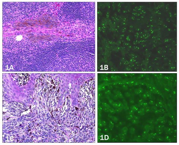
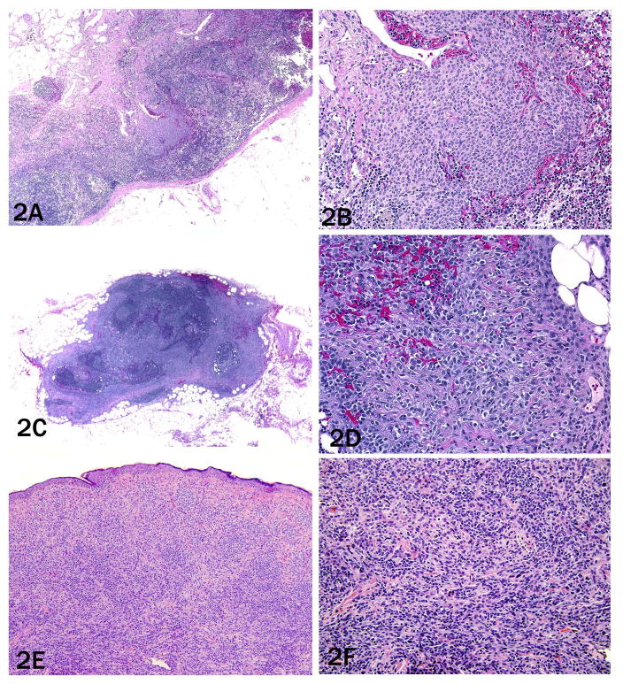
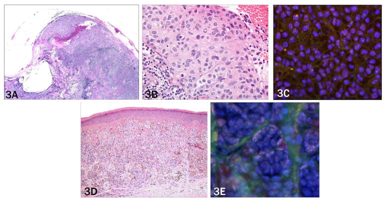
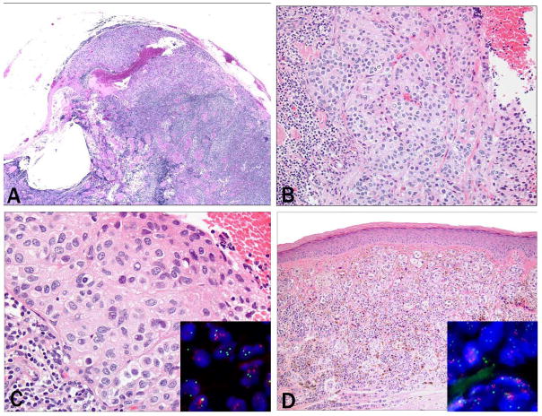
With the increase in sentinel lymph node biopsies in melanoma patients, pathologists are frequently confronted with small deposits of morphologically bland melanocytes in the node, which occasionally cannot be readily classified as benign nodal nevi or melanoma. As most melanomas harbor characteristic chromosomal aberrations which can be used to distinguish them from benign nevi, we used fluorescence in-situ hybridization (FISH) with markers for 3 regions on chromosome 6 and 1 on chromosome 11 to determine the presence of chromosomal aberrations in sentinel lymph node specimens with small foci of melanocytes that had been diagnosed as metastatic melanoma or nodal nevi by histopathology. Fifty-nine tissue samples from 41 patients (24 lymph node metastases, 17 with nodal nevi, and 18 of the available corresponding primary melanomas) were analyzed by FISH. Twenty of 24 (83%) cases diagnosed as metastatic melanoma showed aberrations by FISH. Of the 4 negative cases, 3 were unequivocal melanoma metastases, whereas 1 on re-review was histopathologically equivocal. Of the 17 nodal nevi, 1 (6%) also showed aberrations by FISH, whereas the remainder was negative. Multiple aberrations were present in the positive case, some of which were also found in the corresponding primary tumor, suggesting that this case represents a deceptively bland melanoma metastasis that had been misclassified by histomorphology. Our data indicate that FISH is a useful adjunct tool to traditional methods in the diagnostic workup of deposits of melanocytes in lymph nodes that are histopathologically ambiguous.
Figures
Similar articles
-
Fluorescence in situ hybridization (FISH) analysis of melanocytic nevi and melanomas: sensitivity, specificity, and lack of association with sentinel node status.Int J Surg Pathol. 2012 Oct;20(5):434-40. doi: 10.1177/1066896912445923. Epub 2012 May 4. Int J Surg Pathol. 2012. PMID: 22561674
-
Nevus cell aggregates massively occupying parenchyma of an external iliac lymph node: A case report and review of the literature.J Cutan Pathol. 2020 Dec;47(12):1175-1180. doi: 10.1111/cup.13805. Epub 2020 Sep 10. J Cutan Pathol. 2020. PMID: 32644206 Review.
-
Fatty Acid Synthase and Acetyl-CoA Carboxylase Are Expressed in Nodal Metastatic Melanoma But Not in Benign Intracapsular Nodal Nevi.Am J Dermatopathol. 2018 Apr;40(4):259-264. doi: 10.1097/DAD.0000000000000939. Am J Dermatopathol. 2018. PMID: 28654463 Free PMC article.
-
Diagnostic utility of neural stem and progenitor cell markers nestin and SOX2 in distinguishing nodal melanocytic nevi from metastatic melanomas.Mod Pathol. 2013 Jan;26(1):44-53. doi: 10.1038/modpathol.2012.132. Epub 2012 Aug 17. Mod Pathol. 2013. PMID: 22899289
-
Fluorescence in situ hybridization for ambiguous melanocytic tumors.Histol Histopathol. 2012 Dec;27(12):1539-42. doi: 10.14670/HH-27.1539. Histol Histopathol. 2012. PMID: 23059884 Review.
Cited by
-
From melanocytes to melanomas.Nat Rev Cancer. 2016 Jun;16(6):345-58. doi: 10.1038/nrc.2016.37. Epub 2016 Apr 29. Nat Rev Cancer. 2016. PMID: 27125352 Review.
-
Comparison of melanoma gene expression score with histopathology, fluorescence in situ hybridization, and SNP array for the classification of melanocytic neoplasms.Mod Pathol. 2018 Nov;31(11):1733-1743. doi: 10.1038/s41379-018-0087-6. Epub 2018 Jun 28. Mod Pathol. 2018. PMID: 29955141 Free PMC article.
-
Comparison between melanoma gene expression score and fluorescence in situ hybridization for the classification of melanocytic lesions.Mod Pathol. 2016 Aug;29(8):832-43. doi: 10.1038/modpathol.2016.84. Epub 2016 May 13. Mod Pathol. 2016. PMID: 27174586
-
Assessment of copy number status of chromosomes 6 and 11 by FISH provides independent prognostic information in primary melanoma.Am J Surg Pathol. 2011 Aug;35(8):1146-50. doi: 10.1097/PAS.0b013e318222a634. Am J Surg Pathol. 2011. PMID: 21716079 Free PMC article.
-
Diagnostic and prognostic biomarkers in melanoma.J Clin Aesthet Dermatol. 2014 Jun;7(6):13-24. J Clin Aesthet Dermatol. 2014. PMID: 25013535 Free PMC article. Review.
References
-
- Abrahamsen HN, Hamilton-Dutoit SJ, Larsen J, et al. Sentinel lymph nodes in malignant melanoma: extended histopathologic evaluation improves diagnostic precision. Cancer. 2004;100(8):1683–1691. - PubMed
-
- Bastian BC, Leboit PE, Hamm H, et al. Chromosomal gains and losses in primary cutaneous melanomas detected by comparative genomic hybridization. Cancer Res. 1998;58:2170–5. - PubMed
-
- Biddle DA, Evans HL, Kemp BL, et al. Intraparenchymal nevus cell aggregates in lymph nodes: a possible diagnostic pitfall with malignant melanoma and carcinoma. Am J Surg Pathol. 2003;27(5):673–681. - PubMed
-
- Carson GW, Murray DR, Lyles RH, et al. The amount of metastatic melanoma in a sentinel lymph node: does it have prognostic significance? Ann Surg Oncol. 2003;10(5):575–581. - PubMed
-
- Carson KF, Wen D, Li P, et al. Nodal nevi and cutaneous melanoma. Am J Surg Pathol. 1996;20(7):834–840. - PubMed
MeSH terms
Grants and funding
LinkOut - more resources
Full Text Sources
Other Literature Sources
Medical
Research Materials
