. 2010 Apr;18(4):754-65.
doi: 10.1038/mt.2009.312. Epub 2010 Jan 19.

Intensive pharmacological immunosuppression allows for repetitive liver gene transfer with recombinant adenovirus in nonhuman primates

Affiliations

Intensive pharmacological immunosuppression allows for repetitive liver gene transfer with recombinant adenovirus in nonhuman primates

Antonio Fontanellas et al. Mol Ther. 2010 Apr.

Abstract

Repeated administration of gene therapies is hampered by host immunity toward vectors and transgenes. Attempts to circumvent antivector immunity include pharmacological immunosuppression or alternating different vectors and vector serotypes with the same transgene. Our studies show that B-cell depletion with anti-CD20 monoclonal antibody and concomitant T-cell inhibition with clinically available drugs permits repeated liver gene transfer to a limited number of nonhuman primates with recombinant adenovirus. Adenoviral vector-mediated transfer of the herpes simplex virus type 1 thymidine kinase (HSV1-tk) reporter gene was visualized in vivo with a semiquantitative transgene-specific positron emission tomography (PET) technique, liver immunohistochemistry, and immunoblot for the reporter transgene in needle biopsies. Neutralizing antibody and T cell-mediated responses toward the viral capsids were sequentially monitored and found to be repressed by the drug combinations tested. Repeated liver transfer of the HSV1-tk reporter gene with the same recombinant adenoviral vector was achieved in macaques undergoing a clinically feasible immunosuppressive treatment that ablated humoral and cellular immune responses. This strategy allows measurable gene retransfer to the liver as late as 15 months following the first adenoviral exposure in a macaque, which has undergone a total of four treatments with the same adenoviral vector.


Figures

<b>Figure 1</b>
Figure 1
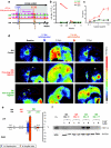
Immunosuppression of macaques with Rituximab and FK506 partially fails in an attempt to transfer twice a transgene to the liver with the same first-generation adenoviral vector. (a) Schematic time line representation of two administrations of a first-generation adenovirus encoding HSV1-tk to three female macaques (individually color-coded) and the immunosuppressive treatments given to two of the animals with Rituximab (20 mg/kg, i.v.) or daily doses of FK506 during the shaded time (1 mg/day). (b) Positron emission tomography (PET) images from the three macaques 2 days after each i.v. administration of AdCMVHSV1-tk performed on the dates marked in a (green arrows) to nonimmunosuppressed 001 control (black), and those animals receiving the immunosuppressive drugs (002-red, 003-green). PET images monitor [18F]FHBG tracer, which becomes phosphorylated and intracellularly retained inside tk-expressing cells. (c) Time course of quantitative analyses of tracer retention in the hepatic region during PET analyses in the animals after the first and second dose of the recombinant AdCMVHS1-tk. Values are normalized by subtraction of the intensity of baseline positron emission in each macaque as measured 1 week before the administration of adenovirus. (d) Follow-up of the titers of neutralizing antiadenoviral antibodies after the first and second administration of AdCMVHSV1-tk to the macaques (color-coded as in a). (e) Double immunostainings of CD20+ and CD19+ cells in peripheral blood mononuclear cell (PBMC) from the indicated color-coded macaques in blood samples drawn upon termination of the second Rituximab course (13 weeks after the first adenoviral administration). (f) In vitro mitogenic responses to adenoviral capsids measured by 3H-Thy incorporation (mean ± SD) of PBMC taken from the indicated macaques (color-coded) 13 weeks after the second adenovirus administration. (g) Fluorescence-activated cell-sorting histograms showing proliferation as estimated on gated CD4+ and CD8+ lymphocytes by CFSE dilution in response to adenoviral capsids at the same time point as in f. Analyses performed in the indicated color-coded macaques. Ad, adenovirus; CFSE, carboxyfluorescein succinimidyl ester; [18F]FHBG, [18F]9-(4-[18F]-fluoro-3-hydroxymethylbutyl)-guanine; HSV1-tk, herpes simplex virus type 1 thymidine kinase; gb, gall bladder; i.v., intravenous; lv, liver; nAb, neutralizing antibody; SI, stimulation index.
<b>Figure 2</b>
Figure 2
An uninterrupted five-drug immunosuppressive regimen (Rituximab+FK506+MMF+ATG+methyl prednisolone) permits efficient liver gene-transduction upon a second administration of an adenoviral vector given 1 month later to an adenovirus naive macaque. This was not feasible in a macaque with signs of previous weak immunity to the viral vector. (a) Schematic representation of the time course of the experiment with the i.v. administrations of AdCMVHSV1.tk that were received by three color-coded macaques and the immunosuppressive regimen given to two of the animals when indicated by the corresponding arrows at the following doses: Rituximab (20 mg/kg), antithymocyte γ-globulin (3 mg/kg), methyl-prednisolone (100 and 50 mg/animal in two daily doses before each adenoviral administration), as well as daily mycophenolate mofetil (30 mg/kg) and FK506 (0.25 mg/kg) during the time indicated by the shaded area. (b) Representation of the pretreatment baseline responsiveness of the color-coded animals to adenoviral capsids as detected by the presence of neutralizing antibodies and (c) PBMC proliferation to serial dilutions of adenoviral capsids performed 2 weeks before the first infusion of AdCMVHSV1-tk. (b) One of the animals (color-coded red) showed low titer antibodies 30 weeks before first adenoviral vector treatment, which became negative in sequential determinations thereafter and (c) low level of antiadenoviral lymphocyte proliferation 10 days before the first administration of adenovirus. (d) Coronal 1 mm-thick positron emission tomography (PET) images from the three macaques 2 days after each intravenous administration of AdCMVHSV1-tk performed on the dates marked in a to nonimmunosuppressed [004 control (black)], and those animals receiving the immunosuppressive drugs [005-red, 006-green]. PET images monitor [18F]FHBG tracer which becomes phosphorylated and intracellularly retained inside tk-expressing cells. Gall bladder (gb) accumulation shows hepatobiliary clearance of the tracer. Dotted lines outline an approximate contour of the liver (lv) based on 3D stacked PET images. (e) Percentage of tk+ cells in ultrasound-guided needle biopsies taken from the right and left liver lobes of the color-coded macaques upon immunohistochemistry stainings. Quantitative data were generated by computer assisted image analyses of 8 non-serial slides counting >1,000 cells. Liver macrophages were distinguished by morphology confirmed with CD68 immunostaining (Supplementary Figure S2c). In the positive macaque a second biopsy performed on day 17 after the second adenoviral administration and showed transgene expression extinction (day +17). (f) Immunoblot analysis of tk and GAPDH (house keeping control) on liver tissue homogenates from the liver biopsies. As a positive control COS7 cells transduced with AdCMVHSV1-tk were used. L and R indicate the hepatic lobe (left and right, respectively) from which the punch biopsy were taken. An additional biopsy was performed 2 weeks after the first set of biopsies (day +17) to assess persistence of expression in the animal successfully retransduced with tk (color-coded green). To corroborate transgene extinction an extra PET study performed one day before that biopsy was negative (images not shown are summarized in Figure 5). Ad, adenovirus; ATG, antithymocyte immunoglobulin; [18F]FHBG, [18F]9-(4-[18F]-fluoro-3-hydroxymethylbutyl)-guanine; i.v., intravenous; MMF, mycophenolate mofetil; nAb, neutralizing antibody; PBMC, peripheral blood mononuclear cell; SI, stimulation index.
<b>Figure 3</b>
Figure 3
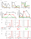
The five drug immunosuppressive regimen lessens humoral and cellular immunity against adenoviral capsid antigens. (a) Follow-up by flow cytometry assessments of the absolute numbers of CD19+ B-lymphocytes, CD4+ T-cells, and CD8+ T-cells in the peripheral blood of the indicated macaques. Arrows point to the dates of AdCMVHSV1-tk injections. (b) Sequential follow-up of serum antiadenoviral neutralizing antibodies in the color-coded animals as described in Figure 2a. (c) Sequential follow-up of the proliferative response of PBMC from the indicated color-coded macaques to adenoviral capsids. (d,e) Proliferation assessed by CFSE dilution in gated (d) CD4+ T-cells and (e) CD8+ T-cells drawn from the peripheral blood of the indicated color-coded macaques 6 weeks after the second adenoviral infusion. CFSE, carboxyfluorescein succinimidyl ester; nAb, neutralizing antibody.
<b>Figure 4</b>
Figure 4
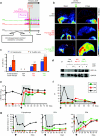
The five-drug regimen permits gene retransfer upon a third readministration of AdCMV-tk given 8 months after the first intravenous adenoviral vector administration. The macaques in the second cohort (005 and 006) were weaned from immunosuppressive treatment 9 weeks after the second administration of AdCMVHSV1-tk and remained 18 weeks off immunosuppressants. (a) Schematic timeline representation of a third administration of AdCMVHSV1-tk to the second cohort of macaques on day 0 and the indicated immunosuppressive regimen given to the color-coded macaques 005 and 006, which had undergone immunosuppression in the previous treatment. (b) Positron emission tomography (PET) images showing liver expression of the transgene before and 2 days after adenoviral administration in the indicated macaques. Of note, the 005 macaque achieved objective gene transfer. (c,d) Immunohistochemical and immunoblot assessment of tk expression in ultrasound-guided liver biopsies from the indicated animals. A biopsy from an additional macaque which had not received adenovirus previously was included as a control in c. Experiments were performed as in Figure 2e,f and biopsies were from the left (L) and right (R) liver lobe as indicated: (e) Follow-up of antiadenovirus neutralizing antibody (nAb) titers in the indicated macaques. (f) PBMC proliferation to adenoviral capsids from the indicated macaques assessed in blood samples drawn at the indicated days following the third AdCMVHSV1-tk administration. (g) Follow-up of the B- and T-cell counts in the peripheral blood of the color-coded macaques in the period of time surrounding the third adenoviral administration. PBMC, peripheral blood mononuclear cell.
<b>Figure 5</b>
Figure 5
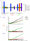
Summary of positron emission tomography (PET) imaging data upon follow-up of the second cohort of macaques undergoing AdCMVHSV1-tk readministrations. (a) Quantitative data from the sequential PET experiments shown in Figure 2d and 4b carried out as indicated again in the graph legend. In these graphs, numeric data represent [18F]FHBG tracer retention measured from 95 to 120 minutes (SUV25) after tracer infusion given separately for the right and left lobe liver areas (upper and lower bars). Results include baseline studies and those performed following the first, second and third AdCMNHSV1-tk administrations. Data include PET studies performed 1 week before the second and third administrations of adenovirus to verify extinction of transgene expression. (b) Time course of quantitative analyses of tracer retention in the hepatic region during the indicated PET analyses in the color-coded animals after each dose of the recombinant AdCMVHS1-tk. Values are normalized by the intensity of baseline positron emission in each macaque as measured 1 week before the administration of adenovirus. [18F]FHBG, [18F]9-(4-[18F]-fluoro-3-hydroxymethylbutyl)-guanine.

Similar articles

Cited by

References

    1. Bessis N, GarciaCozar FJ., and , Boissier MC. Immune responses to gene therapy vectors: influence on vector function and effector mechanisms. Gene Ther. 2004;11 Suppl 1:S10–S17. - PubMed
    1. Bangari DS., and , Mittal SK. Current strategies and future directions for eluding adenoviral vector immunity. Curr Gene Ther. 2006;6:215–226. - PMC - PubMed
    1. Morral N, O'Neal W, Rice K, Leland M, Kaplan J, Piedra PA, et al. Administration of helper-dependent adenoviral vectors and sequential delivery of different vector serotype for long-term liver-directed gene transfer in baboons. Proc Natl Acad Sci USA. 1999;96:12816–12821. - PMC - PubMed
    1. Mingozzi F., and , High KA. Immune responses to AAV in clinical trials. Curr Gene Ther. 2007;7:316–324. - PubMed
    1. Smith TA, White BD, Gardner JM, Kaleko M., and , McClelland A. Transient immunosuppression permits successful repetitive intravenous administration of an adenovirus vector. Gene Ther. 1996;3:496–502. - PubMed

Publication types

MeSH terms

LinkOut - more resources