. 2010 Apr;18(4):692-9.
doi: 10.1038/mt.2009.318. Epub 2010 Jan 19.

Enhancing the therapeutic effect against ovarian cancer through a combination of viral oncolysis and antigen-specific immunotherapy

Affiliations

Enhancing the therapeutic effect against ovarian cancer through a combination of viral oncolysis and antigen-specific immunotherapy

Yu-Qian Zhang et al. Mol Ther. 2010 Apr.

Abstract

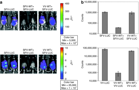
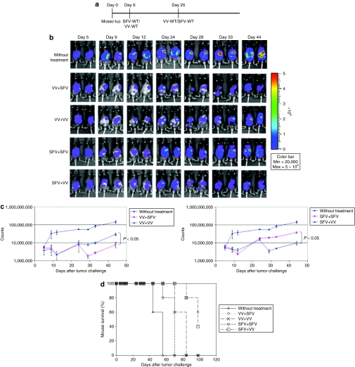
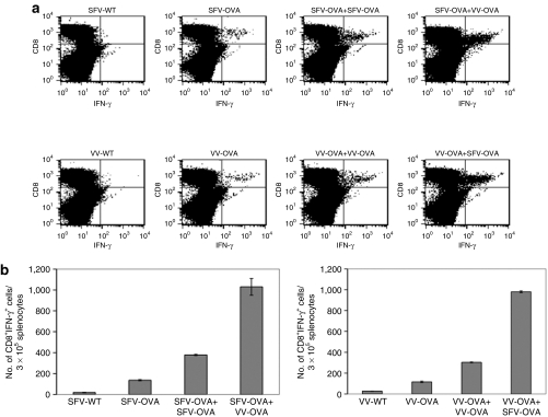
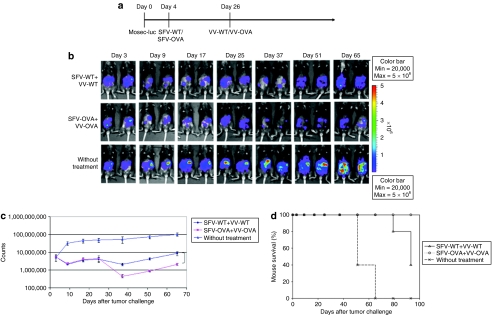
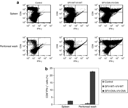
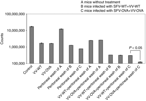
Cancer therapy using oncolytic viruses represents a promising new approach for controlling ovarian cancer. In this study, we have circumvented the limitation of repeated vaccination by employing different virus vectors, Semliki Forest Virus (SFV) and vaccinia virus (VV) for boosting the immune response. We found that infection of tumor-bearing mice with VV followed by infection with SFV or vice versa leads to enhanced antitumor effects against murine ovarian surface epithelial carcinoma (MOSEC) tumors. Furthermore, infection with VV-ovalbumin (OVA) followed by infection with SFV-OVA or vice versa was found to lead to enhanced OVA-specific CD8(+) T-cell immune responses. In addition, we found that infection with SFV-OVA followed by infection with VV-OVA leads to enhanced antitumor effects in vivo and enhanced tumor killing in vitro through a combination of viral oncolysis and antigen-specific immunity. The clinical implications of this study are discussed.


Figures

<b>Figure 1</b>
Figure 1
In vivo luminescence imaging to demonstrate the luciferase expression in tumor-bearing mice infected with SFV or VV. C57BL/6 mice were injected intraperitoneally with 5 × 10⁵/mouse of MOSEC cells on day 0. Mice were then infected with 2 × 10⁶/mouse of SFV-WT or VV-WT on day 3, and the same dose of SFV-luc or VV-luc on day 17. The virus infection was characterized by luciferase expression using luminescence imaging on day 19. (a) Representative luminescence images depicting the fluorescence intensity in tumor-bearing mice infected with SFV/VV. (b) Bar graph depicting the fluorescence intensity in tumor-bearing mice infected with SFV/VV. MOSEC, murine ovarian surface epithelial carcinoma; SFV, Semliki Forest Virus; VV, vaccinia virus; WT, wild type.
<b>Figure 2</b>
Figure 2
In vivo luminescence imaging to demonstrate the antitumor effects generated by infection with SFV or VV in tumor-bearing mice. C57BL/6 mice were injected intraperitoneally with 5 × 10⁵/mouse of MOSEC-luciferase cells on day 0. Mice were then infected with 2 × 10⁶/mouse of SFV-WT or VV-WT on day 6, and the same dose of SFV-WT or VV-WT on day 25. The antitumor effects were characterized by luciferase expression using luminescence imaging. (a) Schematic diagram demonstrating the regimen of infection with SFV or VV. (b) Representative luminescence images depicting the antitumor effects in tumor-bearing mice infected with SFV/VV. (c) Line graphs depicting the fluorescence intensity in tumor-bearing mice infected with SFV/VV. (d) Kaplan–Meier survival analysis of MOSEC-luc tumor-bearing mice infected with SFV/VV. MOSEC, murine ovarian surface epithelial carcinoma; SFV, Semliki Forest Virus; VV, vaccinia virus; WT, wild type.
<b>Figure 3</b>
Figure 3
Infection with SFV-OVA followed by VV-OVA or vice versa leads to enhanced OVA-specific CD8+ T-cell immune responses. C57BL/6 mice were intraperitoneally injected with 5 × 10⁵/mouse of MOSEC cells on day 0. Mice were then infected with 2 × 10⁶ /mouse of SFV-OVA or VV-OVA on day 3, and the same dose of SFV-OVA or VV-OVA on day 17. The control group was injected with a single dose of SFV-WT, VV-WT, SFV-OVA, or VV-OVA on day 3. The spleens were harvested on day 24, the splenocytes were restimulated with OVA peptide SIINFEKL and the OVA-specific T-cell immune responses were characterized using intracellular cytokine staining followed by flow cytometry analysis. (a) Representative flow cytometry data depicting the number of OVA-specific CD8+ T-cells in the various groups. (b) Bar graph representing the number of OVA-specific CD8+ T-cells/3 × 105 splenocytes (mean ± SE). Data shown are representative of two experiments performed. IFN-γ, interferon-γ MOSEC, murine ovarian surface epithelial carcinoma; OVA, ovalbumin; SFV, Semliki Forest Virus; VV, vaccinia virus; WT, wild type.
<b>Figure 4</b>
Figure 4
Infection with SFV-OVA followed by infection with VV-OVA leads to further enhanced antitumor effects. C57BL/6 mice were injected intraperitoneally with 5 × 10⁵/mouse of MOSEC-luciferase cells on day 0. Mice were then infected with 2 × 10⁶/mouse of SFV-WT or SFV-OVA on day 4, and the same dose of VV-WT or VV-OVA on day 26. The antitumor effects were characterized by luminescence imaging. (a) Schematic diagram demonstrating the regimen of infection with SFV or VV. (b) Representative luminescence images depicting the fluorescence intensity in tumor-bearing mice infected with SFV-OVA and VV-OVA compared to SFV-WT and VV-WT. (c) Line graph depicting the fluorescence intensity in tumor-bearing mice infected with SFV-OVA and VV-OVA compared to SFV-WT and VV-WT. (d) Kaplan–Meier survival analysis of MOSEC-luc tumor-bearing mice infected with SFV-OVA and VV-OVA compared to SFV-WT and VV-WT (P = 0.0026). MOSEC, murine ovarian surface epithelial carcinoma; OVA, ovalbumin; SFV, Semliki Forest Virus; VV, vaccinia virus; WT, wild type.
<b>Figure 5</b>
Figure 5
Infection with SFV-OVA followed by VV-OVA leads to enhanced tumor killing by a combination of viral oncolysis and OVA-specific CD8+ T-cells. C57BL/6 mice were intraperitoneally injected with 2 × 10⁶/mouse of MOSEC-luciferase cells on day 0. Mice were then infected with 2 × 10⁶ SFV-WT or SFV-OVA on day 3, and the same dose of VV-WT or VV-OVA on day 13. On day 19, mice were killed, and the peritoneal wash and splenocytes were harvested and the OVA-specific T-cell immune responses were characterized using intracellular cytokine staining followed by flow cytometry analysis. (a) Representative flow cytometry data depicting the number of OVA-specific CD8+ T-cells in the spleens and in the peritoneal wash of infected mice. (b) Bar graph representing the number of OVA-specific CD8+ T-cells/3 × 105 splenocytes (mean ± SE). Data shown are representative of two experiments performed. IFN-γ, interferon-γ MOSEC, murine ovarian surface epithelial carcinoma; OVA, ovalbumin; SFV, Semliki Forest Virus; VV, vaccinia virus; WT, wild type.
<b>Figure 6</b>
Figure 6
In vitro luminescence imaging to demonstrate the cell lysis of MOSEC-luc tumor cells infected with VV-WT or VV-OVA incubated with cells from peritoneal wash of infected mice. MOSEC-luciferase cells were seeded in 24-well plate at 5 × 10⁵ cells/well. A volume of 2 × 10⁶ VV-WT or VV-OVA was added to each well. After 24 hours, the cells were washed with PBS and incubated with 1/8 of the peritoneal wash from the infected mice described in Figure 5 in each well. After another 24 hours, luciferin was added to each well, and the cell lysis was determined by luminescence imaging. Representative bar graph depicting the luciferase expression of MOSEC-luc cells infected with VV-WT or VV-OVA incubated with cells from peritoneal wash of infected mice. MOSEC, murine ovarian surface epithelial carcinoma; OVA, ovalbumin; PBS, phosphate-buffered saline; SFV, Semliki Forest Virus; VV, vaccinia virus; WT, wild type.

Similar articles

Cited by

References

    1. Greenlee RT, Murray T, Bolden S., and , Wingo PA. Cancer statistics, 2000. CA Cancer J Clin. 2000;50:7–33. - PubMed
    1. Jemal A, Siegel R, Ward E, Murray T, Xu J, Smigal C, et al. Cancer statistics, 2006. CA Cancer J Clin. 2006;56:106–130. - PubMed
    1. Baum M, Ebb S., and , Brooks M. Biological fall out from trials of adjuvant tamoxifen in early ovarian cancer. In: Salmon SE (ed.). Adjuvant therapy of cancer V1. Saunders: Philadelphia, pp 269–274; 1990.
    1. Schwartz PE. Current diagnosis and treatment modalities for ovarian cancer. Cancer Treat Res. 2002;107:99–118. - PubMed
    1. Prestwich RJ, Harrington KJ, Pandha HS, Vile RG, Melcher AA., and , Errington F. Oncolytic viruses: a novel form of immunotherapy. Expert Rev Anticancer Ther. 2008;8:1581–1588. - PMC - PubMed

Publication types