Review
. 2010 Apr;30(4):689-702.
doi: 10.1038/jcbfm.2009.282. Epub 2010 Jan 20.

Mast cells as early responders in the regulation of acute blood-brain barrier changes after cerebral ischemia and hemorrhage

Affiliations
Review

Mast cells as early responders in the regulation of acute blood-brain barrier changes after cerebral ischemia and hemorrhage

Perttu Johannes Lindsberg et al. J Cereb Blood Flow Metab. 2010 Apr.

Abstract

The inflammatory response triggered by stroke has been viewed as harmful, focusing on the influx and migration of blood-borne leukocytes, neutrophils, and macrophages. This review hypothesizes that the brain and meninges have their own resident cells that are capable of fast host response, which are well known to mediate immediate reactions such as anaphylaxis, known as mast cells (MCs). We discuss novel research suggesting that by acting rapidly on the cerebral vessels, this cell type has a potentially deleterious role in the very early phase of acute cerebral ischemia and hemorrhage. Mast cells should be recognized as a potent inflammatory cell that, already at the outset of ischemia, is resident within the cerebral microvasculature. By releasing their cytoplasmic granules, which contain a host of vasoactive mediators such as tumor necrosis factor-alpha, histamine, heparin, and proteases, MCs act on the basal membrane, thus promoting blood-brain barrier (BBB) damage, brain edema, prolonged extravasation, and hemorrhage. This makes them a candidate for a new pharmacological target in attempts to even out the inflammatory responses of the neurovascular unit, and to stabilize the BBB after acute stroke.


Figures

Figure 1
Figure 1
Mast cells in human cerebral and meningeal tissues. (A) Antitryptase-stained section of the human cerebral cortex showing predominantly spindle-shaped MCs (arrows) in the characteristic perivascular position. Modified with permission from Ann Med 2009; 41:438–450. (B) A round/oval perivascular mast cell in human cerebral ischemia typically expressing CD117 (arrows). Thin arrows point to the nuclei of endothelial cells. (C) Anti-tryptase staining showing MCs (arrows) in human meningeoma.
Figure 2
Figure 2
Toluidine blue staining of rat brain sections. Toluidine blue staining of ischemic rat brain sections highlighting the metachromatic (deep violet) heparin-containing cytoplasmic granules associated solely with MCs in an intensely stained round/oval MC (arrows). The typical perivascular position along a penetrating cortical artery must be noted. Arrows point to exocytosed MC granules, and triangles highlight endothelial cells.
Figure 3
Figure 3
Chloroacetate esterase (Leder)-stained mast cells in ischemic rat brain. (A) A partially degranulated MC (arrow) located perivascularly; the perivascular edema must be noted. (B) A nondegranulated MC (arrow) within ischemic brain tissue.
Figure 4
Figure 4
Illustration of the components of the blood–brain barrier (left) and the basal lamina (right). Arrows represent the possible involvement of MC-derived mediators in blood–brain barrier and basal lamina damage. ECM, extracellular matrix; ZO, zona occludens; AF6, afadin; 7H6, tight junction-associated phosphoprotein; MMP, matrix metalloproteinase. Modified with permission from Ann Med 2009; 41:438–450.
Figure 5
Figure 5
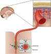
Schematic illustration of the cellular elements of the neurovascular unit. Modified with permission from Ann Med 2009; 41:438–450.

Similar articles

Cited by

References

    1. Abbott NJ. Inflammatory mediators and modulation of blood–brain barrier permeability. Cell Mol Neurobiol. 2000;20:131–147. - PMC - PubMed
    1. Andriopoulou P, Navarro P, Zanetti A, Lampugnani MG, Dejana E. Histamine induces tyrosine phosphorylation of endothelial cell-to-cell adherens junctions. Arterioscler Thromb Vasc Biol. 1999;19:2286–2297. - PubMed
    1. Asahi M, Asahi K, Jung JC, del Zoppo GJ, Fini ME, Lo EH. Role for matrix metalloproteinase 9 after focal cerebral ischemia: effects of gene knockout and enzyme inhibition with BB-94. J Cereb Blood Flow Metab. 2000;20:1681–1689. - PubMed
    1. Asahi M, Wang X, Mori T, Sumii T, Jung JC, Moskowitz MA, Fini ME, Lo EH. Effects of matrix metalloproteinase-9 gene knock-out on the proteolysis of blood-brain barrier and white matter components after cerebral ischemia. J Neurosci. 2001;21:7724–7732. - PMC - PubMed
    1. Asarian L, Yousefzadeh E, Silverman AJ, Silver R. Stimuli from conspecifics influence brain mast cell population in male rats. Horm Behav. 2002;42:1–12. - PMC - PubMed

Publication types

MeSH terms

LinkOut - more resources