. 2010 Jan 19;8(1):e1000281.
doi: 10.1371/journal.pbio.1000281.

The compartmentalized bacteria of the planctomycetes-verrucomicrobia-chlamydiae superphylum have membrane coat-like proteins

Affiliations

The compartmentalized bacteria of the planctomycetes-verrucomicrobia-chlamydiae superphylum have membrane coat-like proteins

Rachel Santarella-Mellwig et al. PLoS Biol. .

Erratum in

Abstract

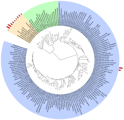
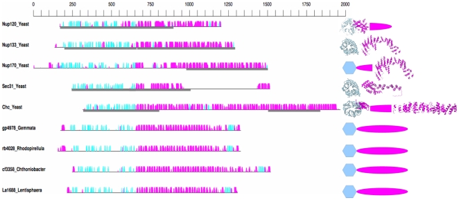
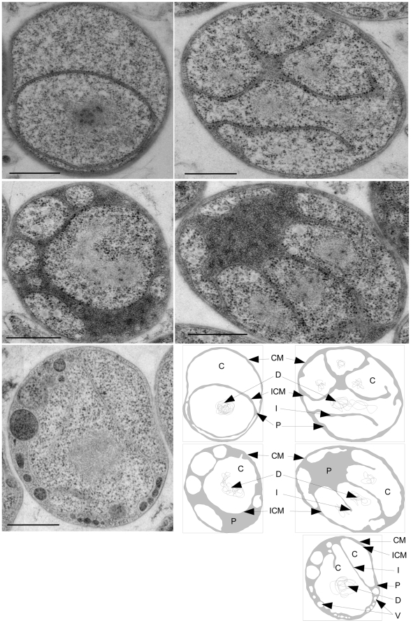
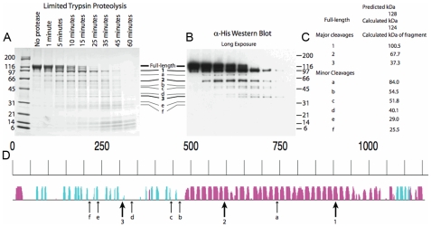
The development of the endomembrane system was a major step in eukaryotic evolution. Membrane coats, which exhibit a unique arrangement of beta-propeller and alpha-helical repeat domains, play key roles in shaping eukaryotic membranes. Such proteins are likely to have been present in the ancestral eukaryote but cannot be detected in prokaryotes using sequence-only searches. We have used a structure-based detection protocol to search all proteomes for proteins with this domain architecture. Apart from the eukaryotes, we identified this protein architecture only in the Planctomycetes-Verrucomicrobia-Chlamydiae (PVC) bacterial superphylum, many members of which share a compartmentalized cell plan. We determined that one such protein is partly localized at the membranes of vesicles formed inside the cells in the planctomycete Gemmata obscuriglobus. Our results demonstrate similarities between bacterial and eukaryotic compartmentalization machinery, suggesting that the bacterial PVC superphylum contributed significantly to eukaryogenesis.


Conflict of interest statement

The authors have declared that no competing interests exist.

Figures

Figure 1
Figure 1. MC architecture detection.
Global phylogeny of 212 organisms for which an alignment of 31 universal protein families could be built, adapted from , drawn with iTOL . Eukaryotes, archaea, and eubacteria are grouped with orange, green, and blue backgrounds, respectively. The number of MC proteins found in each proteome is indicated on the external arc with red bars (see Supporting Information for the complete proteome dataset). Note that this tree includes only two members of the PVC superphylum (both are planctomycetes).
Figure 2
Figure 2. Secondary and tertiary structure of MC proteins.
Representative yeast and PVC MCs are illustrated. Left: predicted secondary structure. The amino-acid scale is represented at the top. The black horizontal line represents the sequence of each MC protein. The predicted secondary structure , α-helices (magenta) and β -strands (cyan) are indicated by colored bars above each line. The height of the bars is proportional to the confidence of the predictions. When an atomic structure is available, the corresponding fragment is highlighted by a grey box below the sequence. Sequences are aligned around the transition from mainly β-sheet to mainly α-helical. Right: predicted and observed tertiary structure: Predicted fold types are represented by coloured shapes, cyan hexagon for β-propeller and magenta oval for SPAH domain. Where known, the atomic structure is represented with the same coloring scheme. PDB codes of the represented structures are 3hxr and 3f7f , Nup120; 1xks and 3i4r , Nup133; 3i5p , Nup170; 1bpo and 1b89 , clathrin; and 2pm6 and 2pm9 , Sec31. Chc, clathrin heavy chain.
Figure 3
Figure 3. The Gemmata membrane morphology is variable.
Electron micrographs of whole sectioned G. obscuriglobus cells representative of the morphologies observed. Lower right: schematic of the electron micrographs with the paryphoplasm colored in grey. CM, cytoplasmic membrane (+cell wall); ICM, intracytoplasmic membrane; P, paryphoplasm; I, invaginations of the ICM; D, DNA; V, vesicle. Scale bar: 500 nm.
Figure 4
Figure 4. Limited proteolysis of gp4978.
Purified N-terminal 10-His tagged gp4978 was trypsin digested and the reaction was stopped at various time points. The resulting fragments were electrophoretically separated. (A) Coomassie-stained SDS-page gel; (B) Anti-His antibodies stained Western blot; (C) Molecular weight of the resulting fragments (the full-length protein has a predicted weight of 128 kDa and a calculated one of 124 kDa); (D) Positions of cleavage are reported on the predicted secondary structure (Figure 2). The size of the arrow is relative to the susceptibility of the positions to cleavage.
Figure 5
Figure 5. Sub-population of gp4978 associates with membranes.
(A, B, and C) Electron micrographs of gp4978-immuno-labelled intra-paryphoplasmic vesicle-bearing G. obscuriglobus cells. Gold particles associated to membranes are indicated by arrows. Scale bars: 500 nm. (D) Chart of the distribution of 494 gold particles in the Cytoplasm (C), Paryphoplasm (P), Vesicle Membranes (VM), Cytoplasmic membrane (CM), and Intracytoplasmic membrane (ICM).

References

    1. Matsuoka K, Orci L, Amherdt M, Bednarek S. Y, Hamamoto S, et al. COPII-coated vesicle formation reconstituted with purified coat proteins and chemically defined liposomes. Cell. 1998;93:263–275. - PubMed
    1. Kirchhausen T. Three ways to make a vesicle. Nat Rev Mol Cell Biol. 2000;1:187–198. - PubMed
    1. Devos D, Dokudovskaya S, Alber F, Williams R, Chait B. T, et al. Components of coated vesicles and nuclear pore complexes share a common molecular architecture. PLoS Biol. 2004;2:e380. doi: 10.1371/journal.pbio.0020380. - DOI - PMC - PubMed
    1. Brohawn S. G, Leksa N. C, Spear E. D, Rajashankar K. R, Schwartz T. U. Structural evidence for common ancestry of the nuclear pore complex and vesicle coats. Science. 2008;322:1369–1373. - PMC - PubMed
    1. Hsia K, Stavropoulos P, Blobel G, Hoelz A. Architecture of a coat for the nuclear pore membrane. Cell. 2007;131:1313–1326. - PMC - PubMed

Publication types