

Biallelic mutation of protocadherin-21 (PCDH21) causes retinal degeneration in humans
- PMID: 20087419
- PMCID: PMC2806159
Biallelic mutation of protocadherin-21 (PCDH21) causes retinal degeneration in humans
Abstract
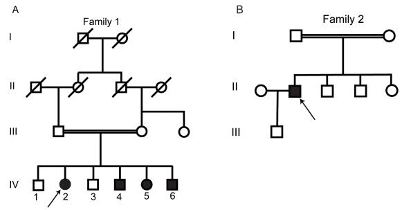
Purpose: To describe the clinical findings and mutations in affected members of two families with an autosomal recessive retinal dystrophy associated with mutations in the protocadherin-21 (PCDH21) gene.
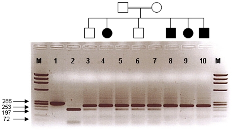
Methods: A full genome scan of members of two consanguineous families segregating an autosomal recessive retinal dystrophy was performed and regions identical by descent identified. Positional candidate genes were identified and sequenced. All patients had a detailed ophthalmic examination, including electroretinography and retinal imaging.
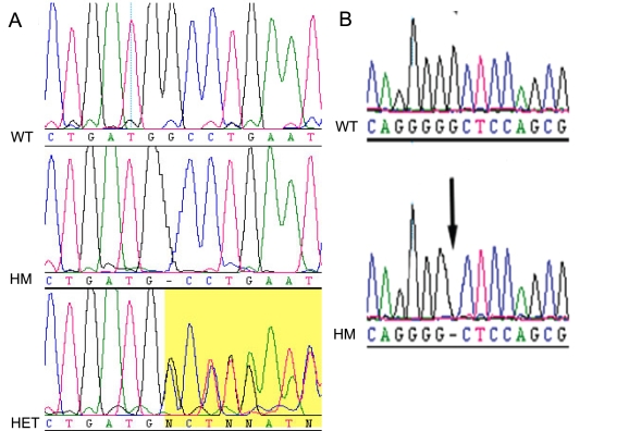
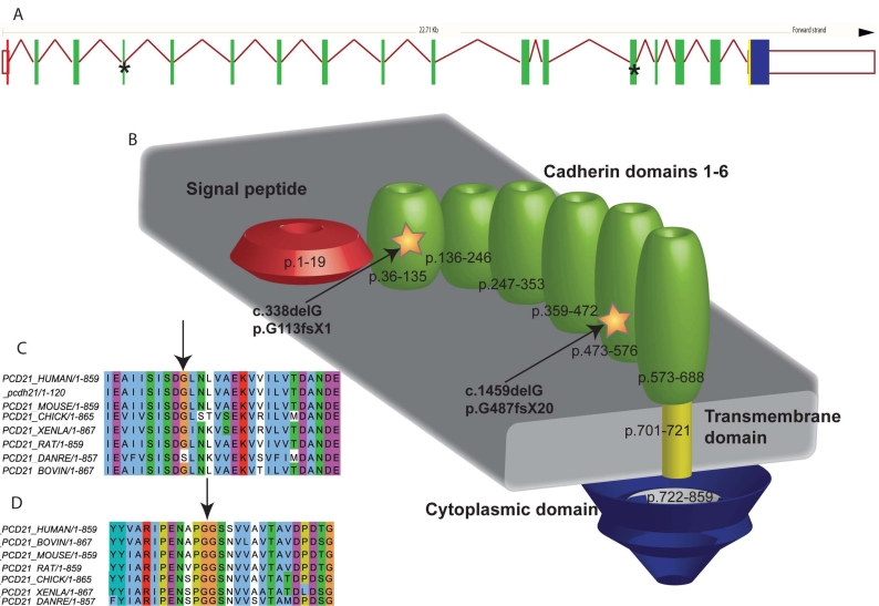
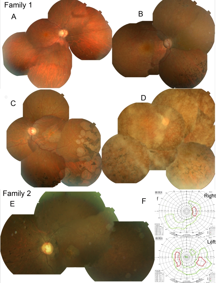
Results: Affected members of both families showed identical homozygosity for an overlapping region of chromosome 10q. Sequencing of a candidate gene, PCDH21, showed two separate homozygous single-base deletions, c.337delG (p.G113AfsX1) and c.1459delG (p.G487GfsX20), which were not detected in 282 control chromosomes. Affected members of the two families first reported nyctalopia in late teenage years and retained good central vision until their late 30s. No color vision was detected in any proband. The fundus appearance included the later development of characteristic circular patches of pigment epithelial atrophy at the macula and in the peripheral retina.
Conclusions: Biallelic mutations in the photoreceptor-specific gene PCDH21 cause recessive retinal degeneration in humans.
Figures
Similar articles
-
Identification of a novel mutation in the CDHR1 gene in a family with recessive retinal degeneration.Arch Ophthalmol. 2012 Oct;130(10):1301-8. doi: 10.1001/archophthalmol.2012.1906. Arch Ophthalmol. 2012. PMID: 23044944 Free PMC article.
-
A novel splice site mutation of CDHR1 in a consanguineous Israeli Christian Arab family segregating autosomal recessive cone-rod dystrophy.Mol Vis. 2012;18:2915-21. Epub 2012 Dec 1. Mol Vis. 2012. PMID: 23233793 Free PMC article.
-
Protocadherin-21 (PCDH21), a candidate gene for human retinal dystrophies.Mol Vis. 2005 Nov 3;11:929-33. Mol Vis. 2005. PMID: 16288196
-
AIPL1 implicated in the pathogenesis of two cases of autosomal recessive retinal degeneration.Mol Vis. 2014 Jan 6;20:1-14. eCollection 2014. Mol Vis. 2014. PMID: 24426771 Free PMC article.
-
Clinical characteristics of recessive retinal degeneration due to mutations in the CDHR1 gene and a review of the literature.Ophthalmic Genet. 2018 Jan-Feb;39(1):51-55. doi: 10.1080/13816810.2017.1363244. Epub 2017 Sep 8. Ophthalmic Genet. 2018. PMID: 28885867 Review.
Cited by
-
A Japanese family with cone-rod dystrophy of delayed onset caused by a compound heterozygous combination of novel CDHR1 frameshift and known missense variants.Hum Genome Var. 2019 Apr 12;6:18. doi: 10.1038/s41439-019-0048-8. eCollection 2019. Hum Genome Var. 2019. PMID: 30992995 Free PMC article.
-
Systematic identification and characterization of novel human skin-associated genes encoding membrane and secreted proteins.PLoS One. 2013 Jun 20;8(6):e63949. doi: 10.1371/journal.pone.0063949. Print 2013. PLoS One. 2013. PMID: 23840300 Free PMC article.
-
INVOLVEMENT OF MULTIPLE MOLECULAR PATHWAYS IN THE GENETICS OF OCULAR REFRACTION AND MYOPIA.Retina. 2018 Jan;38(1):91-101. doi: 10.1097/IAE.0000000000001518. Retina. 2018. PMID: 28406858 Free PMC article. Review.
-
Clinical characteristics of early retinal disease due to CDHR1 mutation.Mol Vis. 2013 Nov 16;19:2250-9. eCollection 2013. Mol Vis. 2013. PMID: 24265541 Free PMC article.
-
Rescue of cone and rod photoreceptor function in a CDHR1-model of age-related retinal degeneration.Mol Ther. 2024 May 1;32(5):1445-1460. doi: 10.1016/j.ymthe.2024.03.026. Epub 2024 Mar 19. Mol Ther. 2024. PMID: 38504520 Free PMC article.
References
-
- Weleber R. Retinitis pigmentosa and allied disorders. In: Ryan SJ, editors. Retina. 2 ed. St.Louis: Mosby; 1994. p. 335–466.
-
- Rattner A, Smallwood PM, Williams J, Cooke C, Savchenko A, Lyubarsky A, Pugh EN, Nathans J. A photoreceptor-specific cadherin is essential for the structural integrity of the outer segment and for photoreceptor survival. Neuron. 2001;32:775–86. - PubMed
-
- Rattner A, Chen J, Nathans J. Proteolytic shedding of the extracellular domain of photoreceptor cadherin. Implications for outer segment assembly. J Biol Chem. 2004;279:42202–10. - PubMed
-
- Bolz H, Ebermann I, Gal A. Protocadherin-21 (PCDH21), a candidate gene for human retinal dystrophies. Mol Vis. 2005;11:929–33. - PubMed
Publication types
MeSH terms
Substances
LinkOut - more resources
Full Text Sources
Molecular Biology Databases