

Recovery from viral encephalomyelitis: immune-mediated noncytolytic virus clearance from neurons
- PMID: 20087684
- PMCID: PMC2891389
- DOI: 10.1007/s12026-009-8143-4
Recovery from viral encephalomyelitis: immune-mediated noncytolytic virus clearance from neurons
Abstract
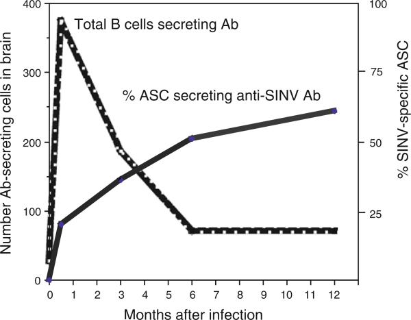
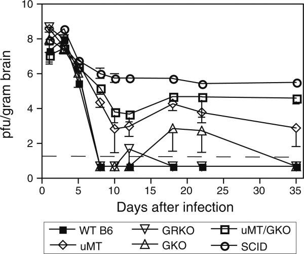
Viral encephalomyelitis is caused by virus infections of neurons in the brain and spinal cord. Recovery is dependent on immune-mediated control and clearance of virus from these terminally differentiated essential cells. Preservation of neuronal function is essential for prevention of neurologic sequelae such as paralysis, seizures and cognitive deficits. Using the model system of Sindbis virus-induced encephalomyelitis in mice, we have shown that immune-mediated clearance of infectious virus from neurons is a noncytolytic process. The major effectors are antibody to the E2 surface glycoprotein produced by B cells, and interferon-gamma produced by T cells. These effectors work in synergy, but neuronal populations differ in their responses to each. Virus is least likely to be cleared from brain neurons and most likely to be cleared from motor neurons in the cervical and thoracic regions of the spinal cord. Because the infected neurons are not eliminated, viral RNA persists and long-term control is needed to prevent virus reactivation. Virus-specific antibody-secreting cells residing in the nervous system after recovery from infection are likely to be important for long-term control.
Figures

Similar articles
-
Interferon-gamma-mediated site-specific clearance of alphavirus from CNS neurons.Science. 2001 Jul 13;293(5528):303-6. doi: 10.1126/science.1059742. Science. 2001. PMID: 11452126
-
The role of antibody in recovery from alphavirus encephalitis.Immunol Rev. 1997 Oct;159:155-61. doi: 10.1111/j.1600-065x.1997.tb01013.x. Immunol Rev. 1997. PMID: 9416509 Review.
-
Synergistic roles of antibody and interferon in noncytolytic clearance of Sindbis virus from different regions of the central nervous system.J Virol. 2007 Jun;81(11):5628-36. doi: 10.1128/JVI.01152-06. Epub 2007 Mar 21. J Virol. 2007. PMID: 17376910 Free PMC article.
-
Germ Line IgM Is Sufficient, but Not Required, for Antibody-Mediated Alphavirus Clearance from the Central Nervous System.J Virol. 2018 Mar 14;92(7):e02081-17. doi: 10.1128/JVI.02081-17. Print 2018 Apr 1. J Virol. 2018. PMID: 29321331 Free PMC article.
-
Role of antibodies in controlling alphavirus infection of neurons.Curr Top Microbiol Immunol. 2001;260:191-200. doi: 10.1007/978-3-662-05783-4_10. Curr Top Microbiol Immunol. 2001. PMID: 11443874 Review. No abstract available.
Cited by
-
TLR7 Is Critical for Anti-Viral Humoral Immunity to EV71 Infection in the Spinal Cord.Front Immunol. 2021 Feb 18;11:614743. doi: 10.3389/fimmu.2020.614743. eCollection 2020. Front Immunol. 2021. PMID: 33679702 Free PMC article.
-
Glucocorticoid treatment of MCMV infected newborn mice attenuates CNS inflammation and limits deficits in cerebellar development.PLoS Pathog. 2013 Mar;9(3):e1003200. doi: 10.1371/journal.ppat.1003200. Epub 2013 Mar 7. PLoS Pathog. 2013. PMID: 23505367 Free PMC article.
-
What Kaplan-Meier survival curves don't tell us about CNS disease.J Neuroimmunol. 2017 Jul 15;308:25-29. doi: 10.1016/j.jneuroim.2017.01.020. Epub 2017 Feb 3. J Neuroimmunol. 2017. PMID: 28187911 Free PMC article.
-
The role of innate versus adaptive immune responses in a mouse model of O'nyong-nyong virus infection.Am J Trop Med Hyg. 2013 Jun;88(6):1170-9. doi: 10.4269/ajtmh.12-0674. Epub 2013 Apr 8. Am J Trop Med Hyg. 2013. PMID: 23568285 Free PMC article.
-
Pre- and post-exposure safety and efficacy of attenuated rabies virus vaccines are enhanced by their expression of IFNγ.Virology. 2015 Jan 1;474:174-80. doi: 10.1016/j.virol.2014.10.025. Epub 2014 Nov 19. Virology. 2015. PMID: 25463615 Free PMC article.
References
-
- Griffin DE. Alphaviruses. In: Knipe DL, Howley PM, Griffin DE, editors. Field's Virology. 5th ed. Lippincott Williams & Wilkins; Philadelphia: 2007.
-
- Center for Disease Control Eastern equine encephalitis—New Hampshire and Massachusetts, August–September 2005. MMWR. 2006;55:697–700. - PubMed
-
- Weaver SC, Salas R, Rico-Hesse R, et al. Re-emergence of epidemic Venezuelan equine encephalomyelitis in South America. VEE study group. Lancet. 1996;348:436–40. - PubMed
-
- Strauss EG, Strauss JH. Structure and replication of the alphavirus genome. In: Schlesinger S, Schlesinger MJ, editors. The togaviridae and flaviviridae. Plenum Publ. Corp; New York: 1986.
Publication types
MeSH terms
Substances
Grants and funding
LinkOut - more resources
Full Text Sources
