

Activity patterns of the diaphragm during voluntary movements in awake cats
- PMID: 20087707
- PMCID: PMC10717415
- DOI: 10.1007/s12576-009-0081-3
Activity patterns of the diaphragm during voluntary movements in awake cats
Abstract
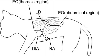
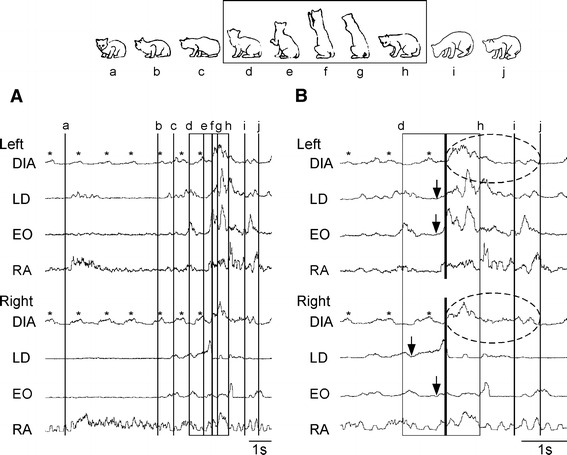
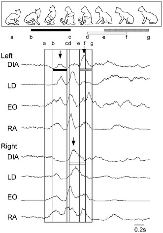
The diaphragm is an important inspiratory muscle, and is also known to participate in the postural function. However, the activity of the diaphragm during voluntary movements has not been fully investigated in awake animals. In order to investigate the diaphragmatic activity during voluntary movements such as extending or rotating their body, we analyzed the electromyogram (EMG) of the diaphragm and trunk muscles in the cat using a technique for simultaneous recordings of EMG signals and video images. Periodic respiratory discharges occurred in the left and right costal diaphragm when the cat kept still. However, once the cat moved, their periodicity and/or synchrony were sometimes buried by non-respiratory activity. Such non-periodic diaphragmatic activities during voluntary movements are considered as the combination of respiratory activity and non-respiratory activity. Most of the diaphragmatic activities started shortly after the initiation of standing-up movements and occurred after the onset of trunk muscle activities. Those activities were more active compared to the normal respiratory activity. During rotation movements, left and right diaphragmatic activities showed asymmetrical discharge patterns and higher discharges than those during the resting situation. This asymmetrical activity may be caused by taking different lengths of each side of the diaphragm and trunk muscles. During reaching movements, the diaphragmatic activity occurred prior to or with the onset of trunk muscle activities. It is likely that diaphragmatic activities during reaching movements and standing-up movements may have been controlled by some different control mechanisms of the central nervous system. This study will suggest that the diaphragmatic activity is regulated not only by the respiratory center but also by inputs from the center for voluntary movements and/or sensory reflex pathways under the awake condition.
Figures

Similar articles
-
The diaphragmatic activities during trunk movements.Adv Exp Med Biol. 2010;669:253-6. doi: 10.1007/978-1-4419-5692-7_51. Adv Exp Med Biol. 2010. PMID: 20217360
-
Postural activity of the diaphragm is reduced in humans when respiratory demand increases.J Physiol. 2001 Dec 15;537(Pt 3):999-1008. doi: 10.1111/j.1469-7793.2001.00999.x. J Physiol. 2001. PMID: 11744772 Free PMC article.
-
The value of extra-diaphragmatic inspiratory muscle surface electromyography during postural control tasks in patients with chronic obstructive pulmonary disease.Respir Med. 2025 Jul;243:108127. doi: 10.1016/j.rmed.2025.108127. Epub 2025 Apr 25. Respir Med. 2025. PMID: 40288657
-
Control of voluntary trunk movements in man. Mechanisms for postural equilibrium during standing.Acta Physiol Scand Suppl. 1990;595:1-60. Acta Physiol Scand Suppl. 1990. PMID: 2080712 Review.
-
Contributions to the understanding of gait control.Dan Med J. 2014 Apr;61(4):B4823. Dan Med J. 2014. PMID: 24814597 Review.
Cited by
-
Electrophysiological properties of Ia excitation and recurrent inhibition in cat abdominal motoneurons.J Physiol Sci. 2019 Mar;69(2):253-262. doi: 10.1007/s12576-018-0643-3. Epub 2018 Oct 15. J Physiol Sci. 2019. PMID: 30324557 Free PMC article.
References
-
- van Lunteren E, Haxhiu MA, Cherniack NS, Goldman MD. Differential costal and crural diaphragm compensation for posture changes. J Appl Physiol. 1985;58:1895–1900. - PubMed
-
- Miyashita M, Suzuki-Inatomi T, Hirai N. Respiratory control during postural changes in anesthetized cats. J Vestib Res. 2003;13:57–64. - PubMed
-
- Duron B, Marlot D. Intercostal and diaphragmatic electrical activity during wakefulness and sleep in normal unrestrained adult cats. Sleep. 1980;3:269–280. - PubMed
-
- Iscoe S. Respiratory and stepping frequencies in conscious exercising cats. J Appl Physiol. 1981;51:835–839. - PubMed
-
- Kawahara K, Kumagai S, Nakazono Y, Miyamoto Y. Coupling between respiratory and stepping rhythms during locomotion in decerebrate cats. J Appl Physiol. 1989;67:110–115. - PubMed
MeSH terms
LinkOut - more resources
Full Text Sources
Miscellaneous
