. 2010 Mar;44(3):313-21.
doi: 10.3109/10715760903494168.

Oxidative damage of mitochondrial DNA in diabetes and its protection by manganese superoxide dismutase

Affiliations

Oxidative damage of mitochondrial DNA in diabetes and its protection by manganese superoxide dismutase

Sally A Madsen-Bouterse et al. Free Radic Res. 2010 Mar.

Abstract

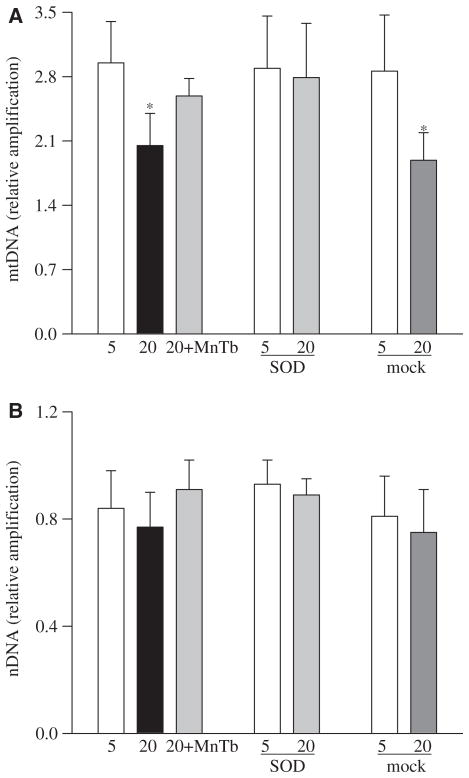
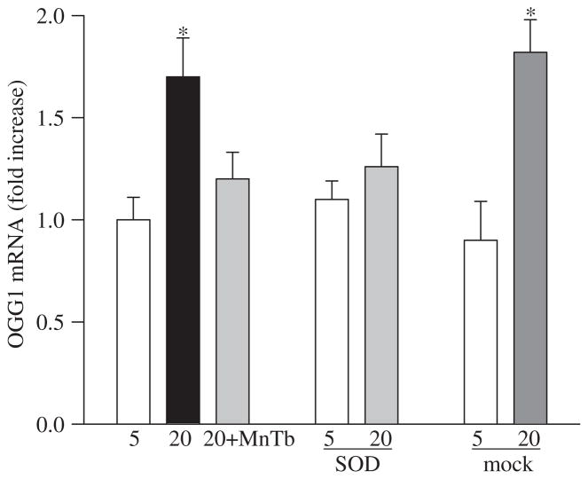
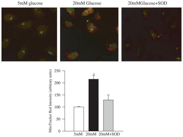
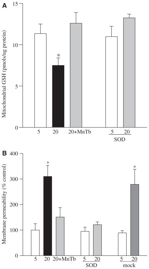
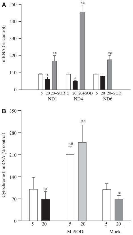
Retinal mitochondria become dysfunctional in diabetes and the production of superoxide radicals is increased; over-expression of MnSOD abrogates mitochondrial dysfunction and prevents the development of diabetic retinopathy. The mitochondrial DNA (mtDNA) is particularly prone to oxidative damage. The aim of this study is to examine the role of MnSOD in the maintenance of mtDNA. The effect of MnSOD mimic, MnTBAP or over-expression of MnSOD on glucose-induced alterations in mtDNA homeostasis and its functional consequence was determined in retinal endothelial cells. Exposure of retinal endothelial cells to high glucose increased mtDNA damage and compromised the DNA repair machinery. The gene expressions of mitochondrial-encoded proteins of the electron transport chain complexes were decreased. Inhibition of superoxide radicals by either MnTBAP or by over-expression of MnSOD prevented mtDNA damage and protected mitochondrial-encoded genes. Thus, the protection of mtDNA from glucose-induced oxidative damage is one of the plausible mechanisms by which MnSOD ameliorates the development of diabetic retinopathy.


Conflict of interest statement

Declaration of interest: This study was supported in part by grants from the National Institutes of Health, Juvenile Diabetes Research Foundation, Thomas Foundation, and Research to Prevent Blindness. The authors report no conflicts of interest. The authors alone are responsible for the content and writing of the paper.

Figures

Figure 1
Figure 1
Effect of high glucose on mitochondrial DNA damage: DNA damage was assessed using 15 ng total DNA and mitochondrial or nuclear specific primers for long and short PCR products in the cells incubated with 5 mM glucose or 20 mM glucose in the presence of MnTBAP or from the cells transfected with MnSOD or Mock cells incubated in 5 mM or 20 mM glucose media. (A) Relative amplification was calculated by normalizing the intensity of the 13.3 kb product to the 232 bp product for mtDNA and (B) the 13.8 kb product to the 241 bp product for nDNA. Each measurement was made in duplicate in at least three different cell preparations. 5=5 mM glucose, 20=20 mM glucose, 20+MnTb=20 mM glucose + 200 μM MnTBAP, SOD=cells transfected with MnSOD, Mock=cells treated with the transfection reagent alone. *p < 0.05 compared to the values obtained from the un-transfected cells incubated in 5 mM glucose.
Figure 2
Figure 2
Effect of glucose on OGG1 and protection by MnSOD:RNA isolated from the cells treated with MnTBAP or transfected with MnSOD or treated with the transfection reagent alone and was assessed by real-time RT-PCR for OGG1. The values were normalized to 18 s mRNA in each sample. Fold-change relative to the values obtained from un-transfected cells incubated in 5 mM glucose was calculated using the ddCt method. Results are from the measurements made in three-to-five preparations. *p < 0.05 compared to control.
Figure 3
Figure 3
Effect of MnSOD on the co-localization of superoxide radicals and OGG1 protein: at the end of incubation in high glucose (4 days) the cells were incubated with Mitotracker Red, washed and fixed with formaldehyde. The cells were permeabilized with Triton X and immunostained for OGG1 using FITC-conjugated secondary antibody. The picture is representative of five different experiments. The accompanying histogram represents the intensity of Mitotracker Red quantified by MetaMorph software in a constant area and constant threshold. 5 mM glucose and 20 mM glucose represent un-transfected cells incubated in 5 mM or 20 mM glucose and 20 mM glucose+SOD=MnSOD transfected cells incubated with 20 mM glucose for 4 days.
Figure 4
Figure 4
Effect of MnSOD on mitochondrial GSH and membrane permeability: mitochondria were prepared from the cells transfected with MnSOD or untransfected, incubated in the presence or absence of MnTBAP. (A) GSH levels were quantified by a colourimetric method and (B) membrane permeability by a spectrophotometric method by measuring the decrease in absorbance at 540 nm induced by calcium chloride.
Figure 5
Figure 5
Effect of glucose on altered abundance of mitochondrial-encoded genes and their protection by MnSOD: transcript abundance was assessed in DNase-treated RNA isolated from retinal endothelial cells using conventional RT-PCR for ND1, ND4 and ND6 of complex I (A) and Cyt b of complex III (B) using the primers provided in Table I. Relative mRNA abundance was quantified using Un-Scan-It Gel digitizing software and the values in the figures are presented as mean band intensity of the target gene normalized by the intensity of β-actin. The values obtained from the un-transfected cells incubated in 5 mM glucose are considered 100% (control). Each measurement was made in four or more different cell preparations. *p < 0.05 compared to control, #p < 0.05 compared to 20 mM glucose alone.

References

    1. Mizutani M, Kern TS, Lorenzi M. Accelerated death of retinal microvascular cells in human and experimental diabetic retinopathy. J Clin Invest. 1996;97:2883–2890. - PMC - PubMed
    1. Kowluru RA, Odenbach S. Effect of long-term administration of alpha lipoic acid on retinal capillary cell death and the development of retinopathy in diabetic rats. Diabetes. 2004;53:3233–3238. - PubMed
    1. Kern TS, Tang J, Mizutani M, Kowluru R, Nagraj R, Lorenzi M. Response of capillary cell death to aminoguanidine predicts the development of retinopathy: comparison of diabetes and galactosemia. Invest Ophthalmol Vis Sci. 2000;41:3972–3978. - PubMed
    1. Kowluru RA, Abbas SN. Diabetes-induced mitochondrial dysfunction in the retina. Inves Ophthalmol Vis Sci. 2003;44:5327–5334. - PubMed
    1. Kowluru RA. Diabetic retinopathy: mitochondrial dysfunction and retinal capillary cell death. Antiox Redox Signal. 2005;7:1581–1587. - PubMed

Publication types

MeSH terms