

Does activation of the FcgammaRIIa play a role in the pathogenesis of the acute lung injury/acute respiratory distress syndrome?
- PMID: 20088831
- PMCID: PMC2811426
- DOI: 10.1042/CS20090422
Does activation of the FcgammaRIIa play a role in the pathogenesis of the acute lung injury/acute respiratory distress syndrome?
Abstract
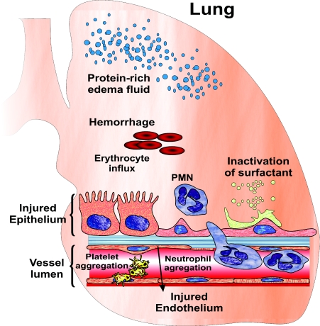
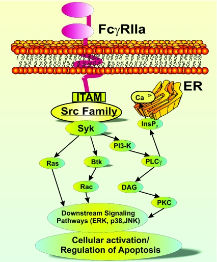
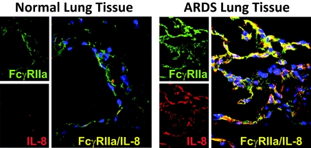
ALI (acute lung injury) and its more severe form ARDS (acute respiratory distress syndrome) are inflammatory diseases of the lung characterized by hypoxaemia and diffuse bilateral infiltrates. Disruption of epithelial integrity and injury to endothelium are contributing factors of the development of ALI/ARDS, and alveolar damage is the most pronounced feature of ALI/ARDS. The resulting increase in lung microvascular permeability promotes influx of inflammatory cells to the alveolar spaces. Oedema fluid contains pro-nflammatory mediators and plasma proteins, including Igs (immunoglobulins). Moreover, several reports describe the presence of autoantibodies and immune complexes [anti-IL-8 (interleukin-8) autoantibody/IL-8 complexes] in lung fluids (oedema and bronchoalveolar lavage fluids) from patients with ALI/ARDS. These immune complexes associate with FcgammaRIIa (Fcgamma IIa receptor) in lungs of patients with ARDS. Furthermore, the expression of FcgammaRIIa is substantially elevated in lungs of these patients. FcgammaRIIa appears on virtually all myeloid cells, platelets and endothelial cells. It is a low-affinity receptor for IgG that preferentially binds aggregated immunoglobulins and immune complexes. FcgammaRs regulate phagocytosis and cell-mediated cytotoxicity, and initiate the release of inflammatory mediators. It should be noted that immune complexes formed between either anti-neutrophil autoantibodies and their specific antigens or anti-HLA (human leucocyte antigen) antibodies and target antigens are implicated in the pathogenesis of TRALI (transfusion-related acute lung injury), and importantly, animal studies indicate that FcgammaRs are essential for these complexes to cause damage to the lungs. Therefore, we hypothesize that FcgammaRs such as FcgammaRIIa could contribute to the pathogenesis of ALI/ARDS.
Figures
Similar articles
-
Anti-chemokine autoantibody:chemokine immune complexes activate endothelial cells via IgG receptors.Am J Respir Cell Mol Biol. 2009 Aug;41(2):155-69. doi: 10.1165/rcmb.2008-0183OC. Epub 2008 Dec 23. Am J Respir Cell Mol Biol. 2009. PMID: 19109244 Free PMC article.
-
Proinflammatory activity of anti-IL-8 autoantibody:IL-8 complexes in alveolar edema fluid from patients with acute lung injury.Am J Physiol Lung Cell Mol Physiol. 2004 Jun;286(6):L1105-13. doi: 10.1152/ajplung.00277.2003. Epub 2004 Jan 16. Am J Physiol Lung Cell Mol Physiol. 2004. PMID: 14729508
-
Anti-interleukin-8 autoantibody:interleukin-8 immune complexes in acute lung injury/acute respiratory distress syndrome.Clin Sci (Lond). 2008 Mar;114(6):403-12. doi: 10.1042/CS20070272. Clin Sci (Lond). 2008. PMID: 18260828 Review.
-
Anti-interleukin 8 autoantibody:interleukin 8 immune complexes visualized by laser confocal microscopy in injured lung.Arch Pathol Lab Med. 2007 Mar;131(3):452-6. doi: 10.5858/2007-131-452-AAICVB. Arch Pathol Lab Med. 2007. PMID: 17516748
-
Interplay between pulmonary epithelial stem cells and innate immune cells contribute to the repair and regeneration of ALI/ARDS.Transl Res. 2024 Oct;272:111-125. doi: 10.1016/j.trsl.2024.05.012. Epub 2024 Jun 17. Transl Res. 2024. PMID: 38897427 Review.
Cited by
-
Fas-deficient mice have impaired alveolar neutrophil recruitment and decreased expression of anti-KC autoantibody:KC complexes in a model of acute lung injury.Respir Res. 2012 Oct 9;13(1):91. doi: 10.1186/1465-9921-13-91. Respir Res. 2012. PMID: 23043753 Free PMC article.
-
Biomarkers in acute lung injury: insights into the pathogenesis of acute lung injury.Crit Care Clin. 2011 Apr;27(2):355-77. doi: 10.1016/j.ccc.2010.12.005. Crit Care Clin. 2011. PMID: 21440206 Free PMC article. Review.
-
The bioactivity of soluble Fas ligand is modulated by key amino acids of its stalk region.PLoS One. 2021 Jun 17;16(6):e0253260. doi: 10.1371/journal.pone.0253260. eCollection 2021. PLoS One. 2021. PMID: 34138914 Free PMC article.
-
Silencing Bruton's tyrosine kinase in alveolar neutrophils protects mice from LPS/immune complex-induced acute lung injury.Am J Physiol Lung Cell Mol Physiol. 2014 Sep 15;307(6):L435-48. doi: 10.1152/ajplung.00234.2013. Epub 2014 Aug 1. Am J Physiol Lung Cell Mol Physiol. 2014. PMID: 25085625 Free PMC article. Clinical Trial.
-
Comparative analysis of the alveolar macrophage proteome in ALI/ARDS patients between the exudative phase and recovery phase.BMC Immunol. 2013 Jun 17;14:25. doi: 10.1186/1471-2172-14-25. BMC Immunol. 2013. PMID: 23773529 Free PMC article.
References
-
- Ware L. B., Matthay M. A. The acute respiratory distress syndrome. N. Engl. J. Med. 2000;342:1334–1346. - PubMed
-
- Ware L. B., Pathophysiology of acute lung injury The acute respiratory distress syndrome. Semin. Respir. Crit. Care Med. 2006;27:337–349. - PubMed
-
- Wheeler A. P., Bernard G. R. Acute lung injury and the acute respiratory distress syndrome: a clinical review. Lancet. 2007;369:1553–1565. - PubMed
-
- Meduri G. U., Kohler G., Headly S., Tolley E., Stentz F., Postlethwaite A. Inflammatory cytokines in the BAL of patients with ARDS. Persistent elevation over time predicts poor outcome. Chest. 1995;108:1303–1314. - PubMed
Publication types
MeSH terms
Substances
Grants and funding
LinkOut - more resources
Full Text Sources
Research Materials