. 2010 Jul;12(7):919-29.
doi: 10.1111/j.1462-5822.2010.01440.x. Epub 2010 Jan 20.

E. coli secreted protein F promotes EPEC invasion of intestinal epithelial cells via an SNX9-dependent mechanism

Affiliations

E. coli secreted protein F promotes EPEC invasion of intestinal epithelial cells via an SNX9-dependent mechanism

Andrew W Weflen et al. Cell Microbiol. 2010 Jul.

Abstract

Enteropathogenic Escherichia coli (EPEC) infection requires the injection of effector proteins into intestinal epithelial cells (IECs) via type 3 secretion. Type 3-secreted effectors can interfere with IEC signalling pathways via specific protein-protein interactions. For example, E. coli secreted protein F (EspF) binds sorting nexin 9 (SNX9), an endocytic regulator, resulting in tubulation of the plasma membrane. Our aim was to determine the mechanism of EspF/SNX9-induced membrane tubulation. Point mutation of the SNX9 lipid binding domains or truncation of the EspF SNX9 binding domains significantly inhibited tubulation, as did inhibition of clathrin coated pit (CCP) assembly. Although characterized as non-invasive, EPEC are known to invade IECs in vitro and in vivo. Indeed, we found significant invasion of Caco-2 cells by EPEC, which, like tubulation, was blocked by pharmacological inhibition of CCPs. Interestingly, however, inhibition of dynamin activity did not prevent tubulation or EPEC invasion, which is in contrast to Salmonella invasion, which requires dynamin activity. Our data also indicate that EPEC invasion is dependent on EspF and its interaction with SNX9. Together, these findings suggest that EspF promotes EPEC invasion of IECs by harnessing the membrane-deforming activity of SNX9.


Figures

Figure 1
Figure 1. Point mutations in either the PX or BAR domain of SNX9 attenuate EspF-induced membrane tubulation
HeLa cells were transiently transfected with pEGFP-EspF and pmCherry-SNX9 and assessed for the formation of membrane tubules by epifluorescence microscopy. Transfection of WT mCherry-SNX9 exhibited extensive membrane tubulation (A). In contrast, expression of SNX9 containing point mutations in either the PX (B), BAR (C), or both domains (D) did not induce membrane tubule formation. In these cells, mCherry-SNX9 and EGFP-EspF co-localized to punctate areas (arrowheads). (E) Quantification of punctate areas per cell from images represented in panels B, C and D. Images are representative of three independent experiments.
Figure 2
Figure 2. SNX9 binding by EspF is not sufficient to induce membrane tubulation
A. Schematic representation of EspF constructs. Wild-type EspF (EspFWT) contains three carboxy-terminal proline-rich repeats (PRR), each of which contains one 7aa SNX9 binding site (gray rectangle). B. HeLa cells cotransfected with pmCherry-SNX9 and either pEGFP-N2, pEGFP-EspF117-131, or pEGFP-EspF1-119 displayed normal cellular distribution of mCherry-SNX9. In cells expressing EGFP-EspF1-166 or EGFP-EspFWT, mCherry-SNX9 was localized to tubular vesicles. C. Quantitation of B. 100 HeLa cells co-expressing mCherry-SNX9 and EGFP constructs were evaluated for the presence or absence of tubular structures over three independent experiments. Expression of EGFPEspF117-131 or EGFP-EspF1-119 did not increase the number of mCherry-SNX9-expressing cells with tubules as compared to expression of the pEGFP-N2 vector. Expression of EGFP-EspF1-166 induced a significant increase (p<0.05) in tubule formation in comparison to EGFP-N2, EGFP-EspF117-131 and EGFP-EspF1-119. The percent of cells exhibiting tubules was significantly higher (p<0.05) in cells expressing EGFP-EspFWT than in all other conditions.
Figure 3
Figure 3. Chlorpromazine and monodansyl cadaverine block EspF/SNX9-induced membrane tubulation
HeLa cells co-transfected with EGFP-EspF and mCherry-SNX9 were incubated with DMEM (Untreated), or DMEM with 0.1% DMSO, 60μM chlorpromazine (CPZ), 80μM dynasore, 10mM methyl-beta-cyclodextrin (MBCD), or 400μM monodansyl cadaverine (MDC). Inhibitor treatment did not affect expression levels of either transgene. Both EGFP-EspF and mCherry-SNX9 localized to tubules in all conditions except treatment with CPZ and MDC, in which case both proteins exhibited a punctate staining pattern. Images are representative of three independent experiments.
Figure 4
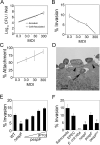
Figure 4. EPEC invasion of Caco-2 intestinal epithelial cells is dependent on EspF/SNX9 interaction
A. Inoculum (○) and total cell-associated bacteria (□) represented as CFU per well of 24-well plates from three independent gentamicin protection assays (GPAs) performed on Caco-2 cells infected with EPEC at MOIs of 0.3, 3, 30 and 300. B. Intracellular CFU per well from the GPAs in A were divided by total cell associated CFU per well and multiplied by 100 to give % invasion. C. Total cell-associated CFU per well were divided by the inoculum and multiplied by 100 to give % attachment. D. Transmission electron microscopic image of Caco-2 cells grown on Transwell permeable supports for 17 days and infected with WT EPEC. The arrow indicates an intracellular bacterium, pedestals are indicated by arrowheads. E. GPAs performed on Caco-2 cells infected with wild-type EPEC (WT), an espF deletion mutant (ΔespF) or ΔespF complemented with the espF gene on an IPTG-inducible plasmid (pespF). Ramp represents treatment of pespF with IPTG concentrations of 0, 1, 10, or 100 μM. F. Invasion of Caco-2 cells by Salmonella (ATCC 14028), WT EPEC, the commensal E. coli isolate HS4, ΔespF, pespF, or pespFD3, an espF point mutant expressing SNX9 binding deficient EspF. All results are representative of three independent experiments. *: p<0.05. **: p<0.05 as compared to*.
Figure 5
Figure 5. Inhibitors of CME and lipid rafts block EPEC invasion but not EPEC-induced TER loss
A. Gentamicin protection assays in Caco-2 cells infected with wild-type EPEC suspended in serum-free DMEM (Untreated) or serum-free DMEM with 0.1% DMSO, 60μM chlorpromazine (CPZ), 80μM dynasore, 10mM methyl-beta-cyclodextrin (MBCD), or 400μM monodansyl cadaverine (MDC). Invasion was significantly inhibited in cells treated with CPZ, MBCD, or MDC. *: p<0.05. Bacterial attachment was not affected by inhibitor treatment (data not shown). Relative invasion was calculated as described in Methods and WT normalized to 1.0. B. GPAs performed as in A but with Salmonella in the presence of media alone (Untreated), 0.1% DMSO or 80μM dynasore. Dynasore treatment significantly reduced the rate of Salmonella invasion. *: p<0.05. C. Caco-2 cells were grown on permeable supports for 14 days and transepithelial electrical resistance (TER) was measured after treatment with serum-free DMEM (Uninfected) or serum-free DMEM with wild-type EPEC (EPEC) for 6 hours. TER was significantly reduced by EPEC infection in the presence of media alone (Untreated) or media with 0.1% DMSO, 60μM chlorpromazine (CPZ), 10mM methyl-beta-cyclodextrin (MBCD), or 400μM monodansyl cadaverine (MDC). Inhibitor treatment alone did not reduce TER. D. HeLa cells transiently transfected with either EGFP-EspF (EspF), YFP-clathrin light chain (Clathrin) or EGFP-dynamin II (Dynamin) were infected for 2.5 hours with wild-type EPEC and processed for epifluorescent microscopy. All three fluorescent fusion proteins were found to accumulate around attached bacteria, shown by DAPI staining (EspF, Clathrin) or by phase contrast (Dynamin).
Figure 6
Figure 6. Depletion of SNX9 by siRNA significantly reduced invasion of Caco-2 cells by EPEC, but not Salmonella
A. Western blot of Caco-2 cells three days after electroporation without oligonucleotide (Mock), with a scrambled RNA oligonucleotide (Scram.), or with SNX9-specific siRNA (siRNA). Image is representative of 6 independent experiments. SNX9 protein levels were assessed in parallel with EPEC and Salmonella invasion assays. B. Integrated density analysis of 6 Western blots represented in A. Values are indicative of SNX9 band density relative to actin band density. *: p<0.05. C. Gentamicin protection assays of Caco-2 cells described in A and infected with WT EPEC at an MOI of 0.3. *: p<0.05. D. Gentamicin protection assays of Caco-2 cells described in A and infected with Salmonella at an MOI of 0.3. *: p<0.05.
Figure 7
Figure 7. Model of SNX9 recruitment and activation during EPEC invasion
A. After injection into the cytosol of epithelial cells, the translocated intimin receptor (Tir) is inserted into the plasma membrane, permitting it to interact with the EPEC surface protein intimin. Tir is known to recruit clathrin, leading to its accumulation at sites of EPEC attachment. B. The combination of EPEC-induced membrane curvature, clathrin assembly and PIP accumulation, all of which are documented to occur upon EPEC attachment, may provide a plasma membrane environment favorable to SNX9 recruitment. Under normal conditions, SNX9 recruits its binding partner dynamin to these membrane domains. C. EspF interacts with the SH3 domain of SNX9 subsequent to its membrane recruitment. EspF then outcompetes with dynamin for interaction with the SNX9 SH3 domain. D. The presence of multiple SNX9 binding domains permits EspF to bind up to three SNX9 molecules, inducing SNX9 oligomerization and increased membrane deforming activity. This, coupled with N-WASP-dependent actin polymerization by EspF, enhances EPEC invasion.

Similar articles

Cited by

References

    1. Abba K, Sinfield R, Hart CA, Garner P. Pathogens associated with persistent diarrhoea in children in low and middle income countries: a systematic review. BMC Infect Dis. 2009;9 - PMC - PubMed
    1. Allen-Vercoe E, Waddell B, Livingstone S, Deans J, DeVinney R. Enteropathogenic Escherichia coli Tir translocation and pedestal formation requires membrane cholesterol in the absence of bundle-forming pili. Cell Microbiol. 2006;8:613–624. - PubMed
    1. Alto NM, Shao F, Lazar CS, Brost RL, Chua G, Mattoo S, et al. Identification of a bacterial type III effector family with G protein mimicry functions. Cell. 2006;124:133–145. - PubMed
    1. Alto NM, Weflen AW, Rardin MJ, Yarar D, Lazar CS, Tonikian R, et al. The type III effector EspF coordinates membrane trafficking by the spatiotemporal activation of two eukaryotic signaling pathways. J Cell Biol. 2007;178:1265–1278. - PMC - PubMed
    1. Andrade JRC, Da Viega VF, De Santa Rosa MR, Suassuna I. An endoctyic process in HEp-2 cells induced by enteropathogenic Escherichia coli. J Med Microbiol. 1989;28:49–57. - PubMed

Publication types

MeSH terms