. 2010 Jun;12(6):844-61.
doi: 10.1111/j.1462-5822.2010.01437.x. Epub 2010 Jan 20.

Qualitative and quantitative ultrastructural analysis of the membrane rearrangements induced by coronavirus

Affiliations

Qualitative and quantitative ultrastructural analysis of the membrane rearrangements induced by coronavirus

Mustafa Ulasli et al. Cell Microbiol. 2010 Jun.

Abstract

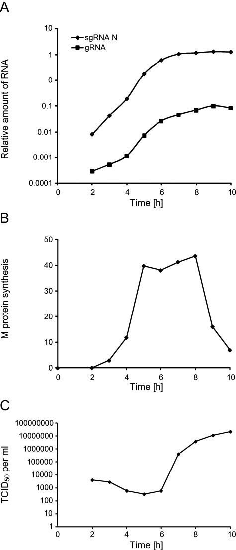
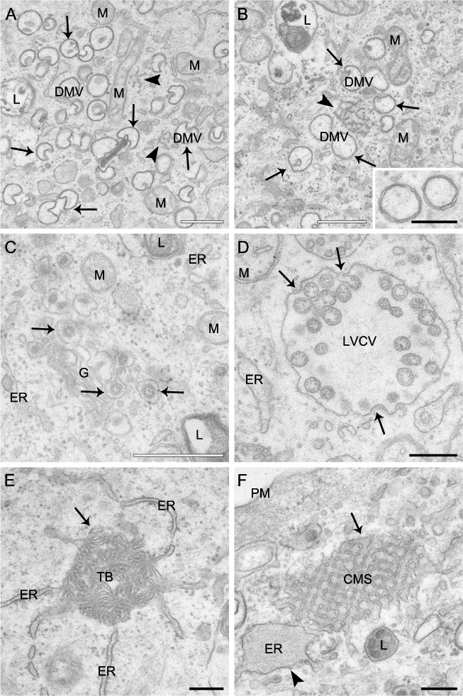
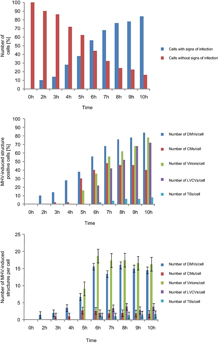
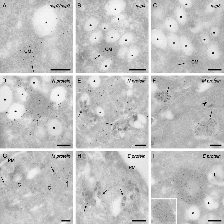
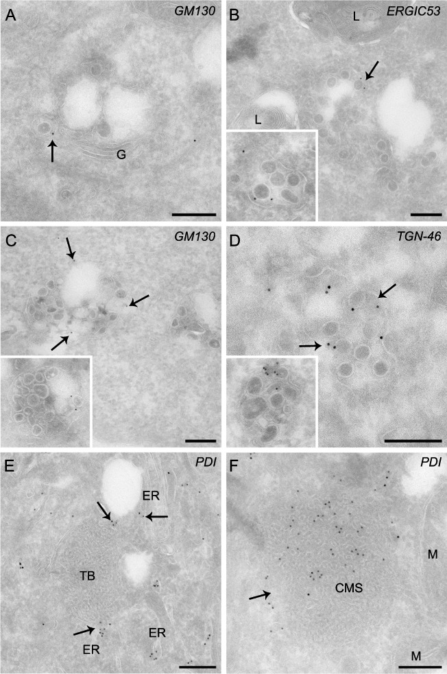
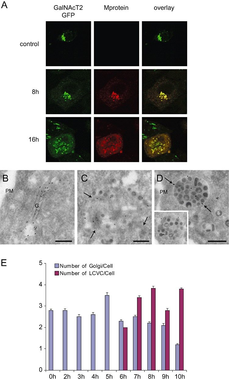
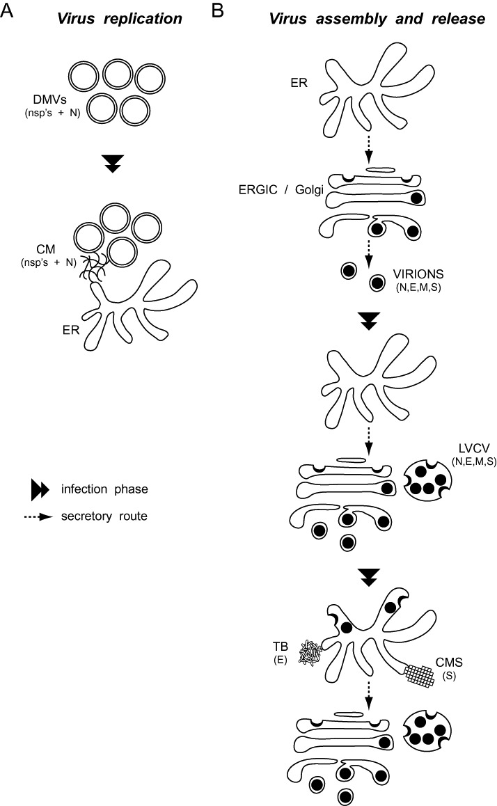
Coronaviruses (CoV) are enveloped positive-strand RNA viruses that induce different membrane rearrangements in infected cells in order to efficiently replicate and assemble. The origin, the protein composition and the function of these structures are not well established. To shed further light on these structures, we have performed a time-course experiment in which the mouse hepatitis virus (MHV)-induced membrane rearrangements were examined qualitatively and quantitatively by (immuno)-electron microscopy. With our approach we were able to confirm the appearance of 6, previously reported, membranous structures during the course of a complete infection cycle. These structures include the well-characterized double-membrane vesicles (DMVs), convoluted membranes (CMs) and virions but also the more enigmatic large virion-containing vacuoles (LVCVs), tubular bodies (TBs) and cubic membrane structures (CMSs). We have characterized the LVCVs, TBs and CMSs, and found that the CoV-induced structures appear in a strict order. By combining these data with quantitative analyses on viral RNA, protein synthesis and virion release, this study generates an integrated molecular and ultrastructural overview of CoV infection. In particular, it provides insights in the role of each CoV-induced structure and reveals that LVCVs are ERGIC/Golgi compartments that expand to accommodate an increasing production of viral particles.


Figures

Figure 1
Figure 1
Analysis of the MHV infection progression by monitoring the viral RNA and protein synthesis, and the extracellular release of viral particles. HeLa‐CEACAM1a cells were infected with MHV‐A59 and the culture supernatants and cell lysates were collected at different p.i. time points as described in Experimental procedures.
A. The total RNA was isolated from the infected cells and the amount of gRNA and sgRNA was quantified by RT‐PCR. Results are expressed using arbitrary units.
B. Infected cells were metabolically labelled 30 min prior of being collected and lysed. Cell lysates were then immunoprecipitated with an antiserum again the complete MHV in combination with anti‐M protein antibodies. Finally, immuno‐complexes were resolved by SDS‐PAGE and the amount of radioactive M protein was quantified. Results are expressed using arbitrary units.
C. The production of the progeny virus was assessed by determining the virus titre of the culture supernatants by end‐point dilutions on LR7 cells and then calculating the TCID50 units per ml of supernatant.
Figure 2
Figure 2
Ultrastructure of membranous structures induced by MHV in host cells. HeLa‐CEACAM1a cells inoculated with MHV‐A59 were fixed at 8 h p.i. and processed for conventional EM as described in Experimental procedures.
A and B. DMVs are cytoplasmic double‐membrane vesicles (arrows) that frequently possess an invagination. DMVs are often found clustered together in close proximity of a small network of membranes, the CMs (arrowheads). The inset in (B) shows a magnification of two DMVs to highlight the two lipid bilayers characterizing these vesicles.
C. Newly made virions (arrows) present in the lumen of the Golgi complex.
D. LVCVs are large circular organelles with a diameter of approximately 450–750 nm that contain numerous virions in their interior. In addition, viral particles can be observed that are assembling at the limiting membrane of this structure by invagination and successive pinching off (arrows).
E. The TBs (arrow) are ball of wool‐like membranous rearrangements with a diameter of approximately 300–650 nm that appear to be continuous with the ER.
F. CMSs are extended (up to 850 nm in length), geometrical and highly organized conformations, which are often seen connected to a swollen ER (arrowhead).
ER, endoplasmic reticulum; G, Golgi complex; M, mitochondria; PM, plasma membrane; L, lysosome. White bar, 500 nm; black bar, 200 nm.
Figure 3
Figure 3
Quantitative analysis of the appearance of the various MHV‐induced structures during the course of an infection. HeLa‐CEACAM1a cells infected with MHV‐A59 were collected at different time points p.i. as described in Experimental procedures before being processed for conventional EM. For the statistical analyses, 50 cell sections were randomly selected for each time point p.i. and used to determine the percentage of cells containing a specific structure (B) and the average number of the same structure per cell section (C). The counted structures were DMVs, CMs, virions, LVCVs and TBs. In addition, the number of cells containing at least one of the six MHV‐induced structures was counted to assess morphologically the proportion of infected cells (A). Error bars in (C) represent the standard deviation of the number of the same structure per cell section.
Figure 4
Figure 4
Non‐structural and structural protein localization in MHV‐infected cells. HeLa‐CEACAM1a cells infected with MHV‐A59 fixed at 10 h p.i. were processed for IEM and immunolabelled as described in Experimental procedures. (A) Nsp2/nsp3, (B) nsp4 and (C) nsp8 are distributed on the limiting membrane of DMVs and on the CMs.
D. The N protein is distributed onto the DMVs and the CMs (arrow).
E. The N nucleocapsid is also present in the viral particles, in this particular image contained in the LVCVs (arrows).
F. The M protein localizes to virions, in this specific case present in the LVCV lumens (arrows), but not to the TBs (arrowhead).
G. The M protein is present in the Golgi complex, both individually or incorporated into luminal virions (arrows).
H. Virions (arrows) are positive for the E protein.
I. The E protein is concentrated into the TBs. The inset shows an additional example.
Arrows point to CMs, asterisks mark the DMVs. M, mitochondria, G, Golgi complex; L, lysosomes; PM, plasma membrane. Bar, 200 nm.
Figure 5
Figure 5
The distribution of various organelle protein markers on MHV‐induced structures. HeLa‐CEACAM1a cells infected with MHV‐A59 were fixed at 10 h p.i., processed for IEM and immunolabelled as described in Experimental procedures.
A. Virions (arrows) present in the Golgi labelled with anti‐GM130 antibodies.
B–D. The LVCVs are positive for ERGIC and Golgi protein markers, e.g. (B) ERGIC53, (C) GM130 and (D) TGN‐46. The insets show additional examples. The arrows highlight the labelling on LVCVs.
E. The ER protein marker PDI localizes to the ER adjacent to the TB but not on this membranous rearrangement.
F. The CMSs (arrow) are derived from the ER because they are positive for PDI.
ER, endoplasmic reticulum; G, Golgi complex; L, lysosomes; M, mitochondria. Bar, 200 nm.
Figure 6
Figure 6
MHV infection leads to morphological alterations of the Golgi complex. HeLa‐GalNAcT2‐GFP cells were inoculated with MHV‐Srec and successively fixed at 0, 8 and 16 h p.i.
A. Golgi viral proteins and virions were detected by IF using antibodies against the M protein for IF. The 16 h p.i. time point was selected because the number of infected cells and the morphological changed induced by MHV are similar to those of HeLa‐CEACAM1a cells infected with MHV‐A59 at 8 h p.i. (not shown).
B–D. The same cells examined in (A) at 0 and 16 h p.i., were also analysed by IEM using anti‐GFP antibodies. A typical Golgi in non‐infected cells is shown in (B). MHV induces the fragmentation of the Golgi cisternae (C, arrows highlight the fragmented cisternae) and the formation of LVCVs (D, arrows), which are positive for the Golgi protein marker GalNAcT2‐GFP. The inset shows an additional example.
E. The EM preparations described in Fig. 2 were used to statistically determine the average number of Golgi, defined as a stack of flattened sacs fragmented or not, and LVCVs per cell profile at the different p.i. time points. Error bars represent standard deviations.
G; Golgi, PM, plasma membrane. Bar, 200 nm.
Figure 7
Figure 7
Schematic representation of the morphological changes induced by CoV in host cells.
A. CoV replication. DMVs are the first membranous rearrangements that CoV induce. Subsequently, CMs become visible in their proximity. CMs appear to be connected with the ER and their size increases with the aggravation of the infection.
B. CoV virion assembly and release. At the early stages of a CoV infection, viral particles principally assemble in the ERGIC and Golgi compartments. An increased synthesis of viral components triggers the expansion of these organelles into LVCVs, which are an additional site where complete virions are produced. A sustained infection and the consequent overflow of the secretory system with viral components results in the assembly of virions in the ER as well, but also in the formation of structures such as the TBs and the CMSs, which are composed by excess of self‐aggregating viral proteins. The composition in viral proteins of each CoV‐induced structure is indicated in between brackets.

References

    1. Ahlquist, P. (2006) Parallels among positive‐strand RNA viruses, reverse‐transcribing viruses and double‐stranded RNA viruses. Nat Rev Microbiol 4: 371–382. - PMC - PubMed
    1. Almsherqi, Z.A. , McLachlan, C.S. , Mossop, P. , Knoops, K. , and Deng, Y. (2005) Direct template matching reveals a host subcellular membrane gyroid cubic structure that is associated with SARS virus. Redox Rep 10: 167–171. - PubMed
    1. Almsherqi, Z.A. , Kohlwein, S.D. , and Deng, Y. (2006) Cubic membranes: a legend beyond the Flatland* of cell membrane organization. J Cell Biol 173: 839–844. - PMC - PubMed
    1. Almsherqi, Z.A. , Landh, T. , Kohlwein, S.D. , and Deng, Y. (2009) Chapter 6: cubic membranes the missing dimension of cell membrane organization. Int Rev Cell Mol Biol 274: 275–342. - PMC - PubMed
    1. Alonso‐Caplen, F.V. , Matsuoka, Y. , Wilcox, G.E. , and Compans, R.W. (1984) Replication and morphogenesis of avian coronavirus in Vero cells and their inhibition by monensin. Virus Res 1: 153–167. - PMC - PubMed

Publication types