. 2010 Jan 20:6:2.
doi: 10.1186/1744-8069-6-2.

Down-regulation of Toll-like receptor 4 gene expression by short interfering RNA attenuates bone cancer pain in a rat model

Affiliations

Down-regulation of Toll-like receptor 4 gene expression by short interfering RNA attenuates bone cancer pain in a rat model

Liu Si Lan et al. Mol Pain. .

Abstract

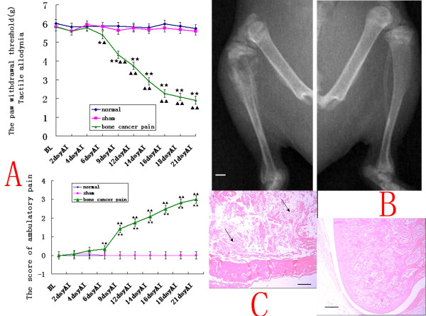
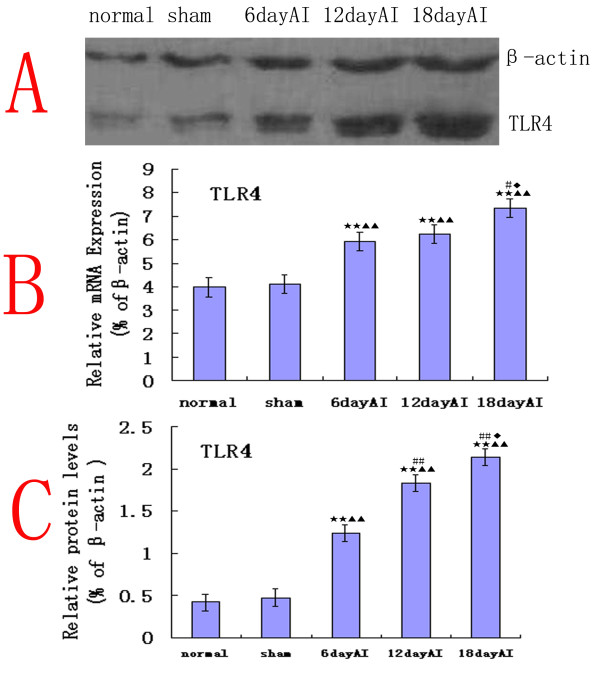
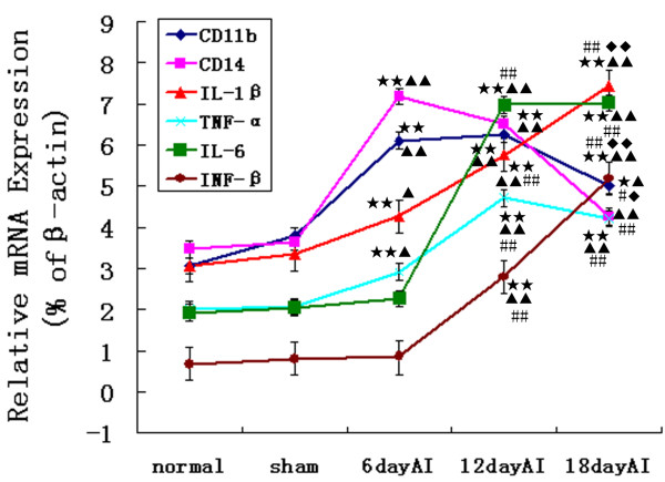
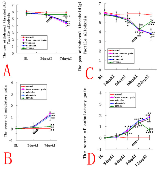
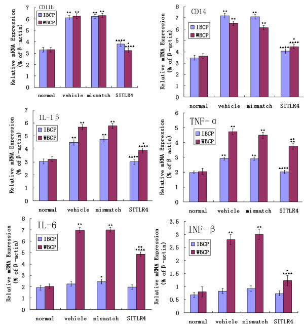
Background: This study demonstrates a critical role in CNS innate immunity of the microglial Toll-like receptor 4 (TLR4) in the induction and maintenance of behavioral hypersensitivity in a rat model of bone cancer pain with the technique of RNA interference (RNAi). We hypothesized that after intramedullary injection of Walker 256 cells (a breast cancer cell line) into the tibia, CNS neuroimmune activation and subsequent cytokine expression are triggered by the stimulation of microglial membrane-bound TLR4.

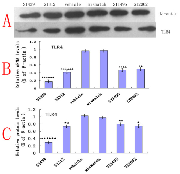
Results: We assessed tactile allodynia and spontaneous pain in female Sprague-Dawley (SD) rats after intramedullary injection of Walker 256 cells into the tibia. In a complementary study, TLR4 small interfering RNA(siRNA) was administered intrathecally to bone cancer pain rats to reduce the expression of spinal TLR4. The bone cancer pain rats treated with TLR4 siRNA displayed significantly attenuated behavioral hypersensitivity and decreased expression of spinal microglial markers and proinflammatory cytokines compared with controls. Only intrathecal injection of TRL4 siRNA at post-inoculation day 4 could prevent initial development of bone cancer pain; intrathecal injection of TRL4 siRNA at post-inoculation day 9 could attenuate, but not completely block, well-established bone cancer pain.

Conclusions: TLR4 might be the main mediator in the induction of bone cancer pain. Further study of this early, specific, and innate CNS/microglial response, and how it leads to sustained glial/neuronal hypersensitivity, might lead to new therapies for the prevention and treatment of bone cancer pain syndromes.


Figures

Figure 1
Figure 1
A rat model of bone pain from metastatic bone cancer. (A) Rats with tibia tumors after Walker 256 cells inoculation displayed both tactile allodynia and spontaneous pain. The PWL progressively decreased and the ambulatory score progressively increased on days 6 (n = 12, ANOVA2w, P < 0.05 pos-hoc Bonferroni, df = 2, F = 1.07), 9, 12, 14, 16, 18, and 21 (ANOVA2w, P < 0.01, df = 2, F = 1.13) after inoculation (AI) in the tumor-bearing group compared with sham-injected controls and normal rats. Results are given as means ± S.D, *p < 0.05 **P < 0.01 vs. normal group; p < 0.05 ▲ ▲p < 0.01 vs. sham group. (B) Radiographs of the left tibiae 18 days after inoculation with Walker 256 cells and the right normal tibiae. There was bilateral cortical bone damage and large bone defects at the 18 days' tumor-bearing proximal epiphysis of the tibiae.(C) HE staining of normal (upper panel) and 18 days of tumor-bearing (lower) proximal epiphysis of the tibiae. Note that tumor cells were densely packed in the marrow cavity and induced the destruction of trabeculae. Arrows indicate tumor cells. Bar = 1 mm in B and 40 μm in C.
Figure 2
Figure 2
TLR4 expression increases in the bone cancer pain model. (B) Real-time quantitative RT-PCR analyses of mRNA temporal expression of TLR4 in rat lumbar spinal cord. The relative levels of spinal TLR4 mRNA significantly increased on days 6, 12 and18 (n = 4, ANOVA1w, P < 0.01, post hoc Dunnett testing, df = 3, F = 1.17) after inoculation (AI) in bone cancer pain rats compared with sham-injected controls and normal rats. The level of gene expression was calculated after normalizing against β-actin in each sample and is presented as relative mRNA expression units. (A, C) The protein level of TLR4 detected by western blotting analysis in spinal cord. A significant up-regulation of TLR4 protein level (n = 4, ANOVA1w, P < 0.01, post hoc Dunnett testing, df = 3, F = 1.06) on the sixth day AI. The protein levels further increased at 12 and 18 days (P < 0.01) after inoculation compared with sham-injected controls and normal rats, respectively. Each data point represents at least three independent experiments. Values are presented as mean ± SEM. *P < 0.05, **P < 0.01 vs. normal group; P < 0.05, ▲▲P < 0.01 vs. sham group; #P < 0.05, ##P < 0.01 vs. 6 day AI; P < 0.05, ◆◆P < 0.01 vs. 12 day AI.
Figure 3
Figure 3
Microglial activation and proinflammatory cytokine expression increases in the bone cancer pain model. Real-time quantitative RT-PCR analyses of the temporal expression of microglial activation markers (CD11b, CD14) and proinflammatory cytokines (IL-6, IL-1β, TNF-α, and INF-β) mRNA in rat lumbar spinal cord. The mRNA expression levels of CD11b and CD14 significantly increased in the bone cancer pain group at six days AI and 12 days AI (n = 4, ANOVA1w, P < 0.01, post hoc Dunnett testing, df = 3, F = 0.98). CD11b mRNA expression was also higher at 18 days AI (P < 0.05). The bone cancer pain group also showed significant up-regulation IL-6, IL-1β, TNF-α, and INF-β mRNA relative to the normal and sham group(n = 4, ANOVA1w, P < 0.01, post hoc Dunnett testing, df = 3, F = 1.11), starting at day six after inoculation for IL-1β and TNF-α (P < 0.05), and at day 12 after inoculation for IL-6 and INF-β (P < 0.01). Values are presented as mean ± SEM. *P < 0.05, **P < 0.01 vs. normal group; P < 0.05, ▲▲ P < 0.01 vs. sham group; #P < 0.05, ##P < 0.01 vs. 6 days AI; P < 0.05, ◆◆P < 0.01 vs. 12 days AI.
Figure 4
Figure 4
Molecular down-regulation of TLR4 mRNA and protein upon treatment with siRNA. (B) TLR4 mRNA levels in microglial cells line (HAPI), normalized to β-actin, were significantly reduced in TLR4 siRNA (SITLR4) versus mismatch siRNA- and vehicle-treated cells 24 h after transfection. siRNA439 was the most effective at knocking down TLR4 expression(n = 4, ANOVA1w, P < 0.001, post hoc Dunnett testing, df = 3, F = 1.08). (A) The TLR4 protein levels detected by western blotting analysis in HAPI 48 h after transfection, normalized to β-actin. (C) TRL4 protein levels were significantly reduced following treatment with TLR4 siRNA (SITLR4) compared with vehicle or mismatch siRNA treatment. Each data point represents at least three independent experiments. Values are presented as mean ± SEM. *P < 0.05, **P < 0.01, ***P < 0.001 vs. vehicle-treated cells; P < 0.05, ▲▲P < 0.01, ▲▲P < 0.001 vs. mismatch siRNA-treated group.
Figure 5
Figure 5
Intrathecal TLR4 siRNA439 attenuates bone cancer pain in the rat model. (A, B) In the siRNA-treated IBCP group, the PWTs were significantly higher and the ambulatory scores were significantly lower than those observed in mismatch siRNA- and vehicle-treated rats(n = 10, ANOVA2w, P < 0.01, pos-hoc Bonferroni, df = 4, F = 1.02), and there were no significant differences compared to normal rats. This indicated that intrathecal injection of TLR4 siRNA439 (SITLR4) could prevent the initial development of bone cancer pain. (C, D) In the siRNA-treated WBCP group, the PWTs were significantly elevated compared with the mismatch siRNA- and vehicle-treated rats(n = 10, ANOVA2w, P < 0.01, pos-hoc Bonferroni, df = 4, F = 0.96), but still lower than in normal rats (P < 0.05) Meanwhile, spontaneous pain was attenuated compared with mismatch siRNA- and vehicle-treated rats(n = 10, ANOVA2w, P < 0.01, pos-hoc Bonferroni, df = 4, F = 1.05). However, the ambulatory score was still higher than in normal rats (P < 0.05), which indicated that intrathecal injection of TLR4 siRNA439 (SITLR4) could alleviate, but not reverse, well-established bone cancer pain. Values are presented as mean ± SEM. *P < 0.05, **P < 0.01 vs. normal group; P < 0.05, P < 0.01 vs. vehicle-treated group; #P < 0.05, ##P < 0.01 vs. bone cancer pain group; P < 0.05, ◆◆P < 0.01 vs. mismatch siRNA-treated group. Arrows indicate the siRNA injection times.
Figure 6
Figure 6
Attenuation of TLR4 expression by Intrathecal TLR4 siRNA439. (A, B) The TLR4 mRNA levels in the L4~6 lumbar spinal cord from mismatch siRNA- and vehicle- treated bone cancer pain rats were higher in IBCP group (A) and in WBCP group (B) (n = 4, ANOVA1w, P < 0.01, post hoc Dunnett testing, df = 3, F = 1.12)relative to that of normal rats. TLR4 siRNA439 (SITLR4) treatment caused a significant decrease in IBCP group, and in WBCP group in the TLR4 mRNA level compared with that in normal rats (P < 0.01). (C, D, E, F) Data from several western blotting experiments revealed a significant up-regulation of TLR4 (n = 4, ANOVA1w, P < 0.01, post hoc Dunnett testing, df = 3, F = 1.16) protein in mismatch siRNA-and vehicle- treated bone cancer pain rats relative to that from normal rats, whether in the IBCP group (C, E) or in the WBCP group (D, F). TLR4 siRNA439 treatment caused a significant decrease (n = 4, ANOVA1w, P < 0.01, post hoc Dunnett testing, df = 3, F = 0.96)relative the level in mismatch siRNA-and vehicle- treated bone cancer pain rats, and a significant decline compared to normal rats (P < 0.05) in the IBCP group (C, E). Values are presented as mean ± SEM. *P < 0.05, **P < 0.01 vs. normal group; P < 0.05, ▲▲ P < 0.01 vs. vehicle-treated group; P < 0.05, ◆◆P < 0.01 vs. mismatch siRNA-treated group.
Figure 7
Figure 7
Decreased bone cancer pain-induced microglial activation and proinflammatory cytokine expression by Intrathecal TLR4 siRNA439. In both IBCP group and WBCP group, Real-time quantitative RT-PCR analyses revealed a significant down-regulation of two microglial activation markers, CD14 and CD11b compared with mismatch siRNA- and vehicle-treated rats(n = 4, ANOVA1w, P < 0.01, post hoc Dunnett testing, df = 3, F = 1.08). Proinflammatory cytokines mRNA expression was also significantly down-regulated, i.e., IL-1β (P < 0.01), TNF-α (P < 0.05), IL-6 (P < 0.01), and INF-β (P < 0.01) in the WBCP group; IL-1β (P < 0.01), and TNF-α (P < 0.01) in the IBCP group compared with rats treated with vehicle or the mismatch siRNA. Values are presented as mean ± SEM. *P < 0.05, **P < 0.01 vs. normal group; P < 0.05, ▲▲ P < 0.01 vs. vehicle-treated group; P < 0.05, ◆◆P < 0.01 vs. mismatch siRNA-treated group.

Similar articles

Cited by

References

    1. Urch C. The pathophysiology of cancer-induced bone pain: current understanding. Palliat Med. 2004;18:267–274. doi: 10.1191/0269216304pm887ra. - DOI - PubMed
    1. Strang P. Cancer pain-a provoker of emotional, social and existential distress. Acta Oncol. 1998;37:641–644. doi: 10.1080/028418698429973. - DOI - PubMed
    1. Mao-Ying Q-L, Zhao J, Dong Z-Q, Wang J, Yu J, Yan M-F, Zhang Y-Q, Wu G-C, Wang Y-Q. A rat model of bone cancer pain induced by intra-tibia inoculation of Walker 256 mammary gland carcinoma cells. Biochem Biophys Res Commun. 2006;345(4):1292–1298. doi: 10.1016/j.bbrc.2006.04.186. - DOI - PubMed
    1. Yao M, Yang JP, Wang LN, Cheng H, Zhang YB, Xu QN, Wu YW. Feasibility of establishment of rat model of bone cancer pain by using Walker 256 cells cultured in vitro or in vivo. Zhonghua Yi Xue Za Zhi. 2008;88(13):880–884. - PubMed
    1. Schwei MJ, Honore P, Rogers SD, Salak-Johnson JL, Finke MP, Ramnaraine ML, Clohisy DR, Mantyh PW. Neurochemical and cellular reorganization of the spinal cord in a murine model of bone cancer pain. J Neurosci. 1999;19(24):10886–10897. - PMC - PubMed

Publication types

MeSH terms