

Radiographic assessment of the femorotibial joint of the CCLT rabbit experimental model of osteoarthritis
- PMID: 20089151
- PMCID: PMC2828401
- DOI: 10.1186/1471-2342-10-3
Radiographic assessment of the femorotibial joint of the CCLT rabbit experimental model of osteoarthritis
Abstract
Background: The purposes of the study were to determine the relevance and validity of in vivo non-invasive radiographic assessment of the CCLT (Cranial Cruciate Ligament Transection) rabbit model of osteoarthritis (OA) and to estimate the pertinence, reliability and reproducibility of a radiographic OA (ROA) grading scale and associated radiographic atlas.
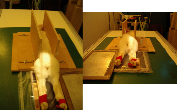
Methods: In vivo non-invasive extended non weight-bearing radiography of the rabbit femorotibial joint was standardized. Two hundred and fifty radiographs from control and CCLT rabbits up to five months after surgery were reviewed by three readers. They subsequently constructed an original semi-quantitative grading scale as well as an illustrative atlas of individual ROA feature for the medial compartment. To measure agreements, five readers independently scored the same radiographic sample using this atlas and three of them performed a second reading. To evaluate the pertinence of the ROA grading scale, ROA results were compared with gross examination in forty operated and ten control rabbits.
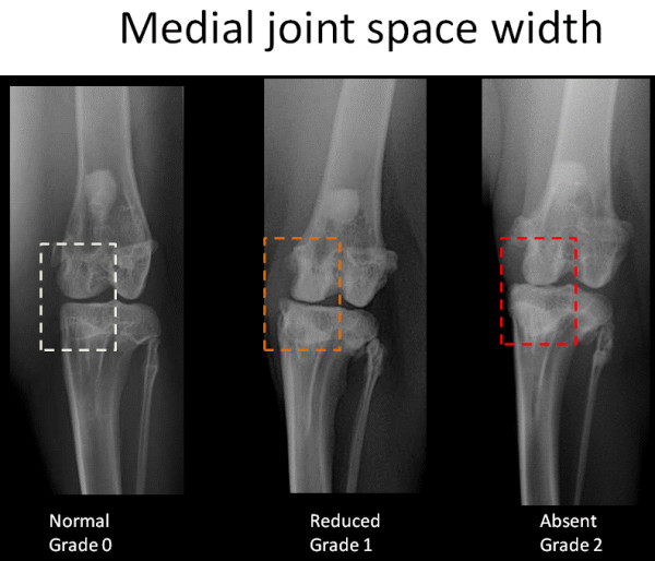
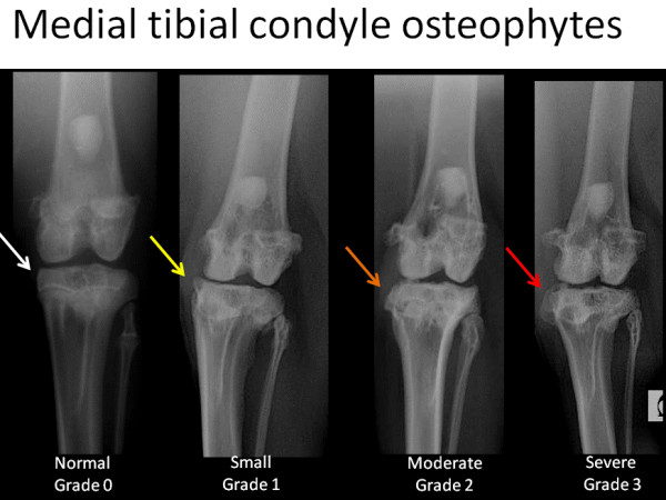
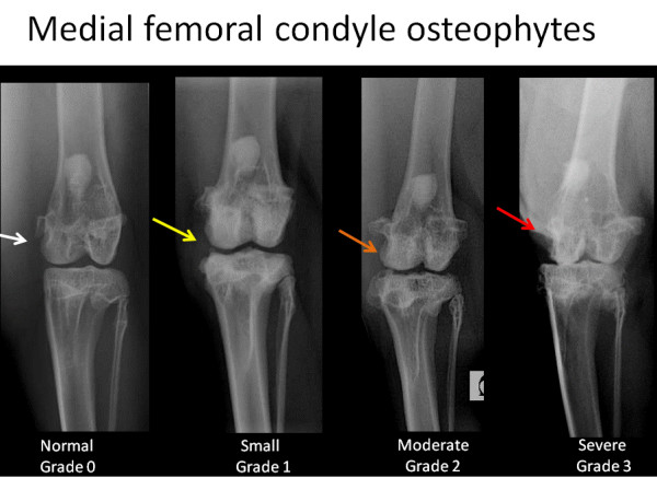
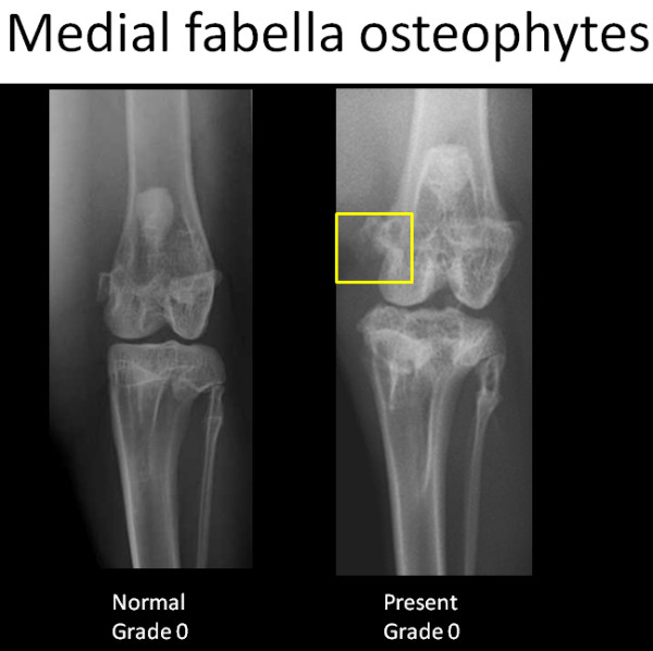
Results: Radiographic osteophytes of medial femoral condyles and medial tibial condyles were scored on a four point scale and dichotomously for osteophytes of medial fabella. Medial joint space width was scored as normal, reduced or absent. Each ROA features was well correlated with gross examination (p < 0.001). ICCs of each ROA features demonstrated excellent agreement between readers and within reading. Global ROA score gave the highest ICCs value for between (ICC 0.93; CI 0.90-0.96) and within (ICC ranged from 0.94 to 0.96) observer agreements. Among all individual ROA features, medial joint space width scoring gave the highest overall reliability and reproducibility and was correlated with both meniscal and cartilage macroscopic lesions (rs = 0.68 and rs = 0.58, p < 0.001 respectively). Radiographic osteophytes of the medial femoral condyle gave the lowest agreements while being well correlated with the macroscopic osteophytes (rs = 0.64, p < 0.001).
Conclusion: Non-invasive in vivo radiography of the rabbit femorotibial joint is feasible, relevant and allows a reproducible grading of experimentally induced OA lesion. The radiographic grading scale and atlas presented could be used as a template for in vivo non invasive grading of ROA in preclinical studies and could allow future comparisons between studies.
Figures
Similar articles
-
Multirater agreement for grading the femoral and tibial cartilage surface lesions at CT arthrography and analysis of causes of disagreement.Eur J Radiol. 2017 Mar;88:95-101. doi: 10.1016/j.ejrad.2016.12.026. Epub 2016 Dec 31. Eur J Radiol. 2017. PMID: 28189216
-
Three-Dimensional Quantitative Morphometric Analysis (QMA) for In Situ Joint and Tissue Assessment of Osteoarthritis in a Preclinical Rabbit Disease Model.PLoS One. 2016 Jan 25;11(1):e0147564. doi: 10.1371/journal.pone.0147564. eCollection 2016. PLoS One. 2016. PMID: 26808542 Free PMC article.
-
Longitudinal (one-year) change in cartilage thickness in knees with early knee osteoarthritis: A within-person between-knee comparison.Arthritis Care Res (Hoboken). 2014 Apr;66(4):636-41. doi: 10.1002/acr.22172. Arthritis Care Res (Hoboken). 2014. PMID: 24106150
-
[Reproducibility of radiologic diagnosis in gonarthrosis].Z Orthop Ihre Grenzgeb. 1997 May-Jun;135(3):197-202. doi: 10.1055/s-2008-1039580. Z Orthop Ihre Grenzgeb. 1997. PMID: 9334072 German.
-
Which radiographic techniques should we use for research and clinical practice?Best Pract Res Clin Rheumatol. 2006 Feb;20(1):39-55. doi: 10.1016/j.berh.2005.08.002. Best Pract Res Clin Rheumatol. 2006. PMID: 16483906 Review.
Cited by
-
RANKL synthesized by articular chondrocytes contributes to juxta-articular bone loss in chronic arthritis.Arthritis Res Ther. 2012 Jun 18;14(3):R149. doi: 10.1186/ar3884. Arthritis Res Ther. 2012. PMID: 22709525 Free PMC article.
-
Clinical magnetic resonance-enabled characterization of mono-iodoacetate-induced osteoarthritis in a large animal species.PLoS One. 2018 Aug 3;13(8):e0201673. doi: 10.1371/journal.pone.0201673. eCollection 2018. PLoS One. 2018. PMID: 30075007 Free PMC article.
-
Use of hyaluronic acid associated with triamcinolone acetonide or ozone gas in the treatment of induced osteoarthritis in rabbits.Acta Cir Bras. 2021 Dec 17;36(12):e361201. doi: 10.1590/ACB361201. eCollection 2021. Acta Cir Bras. 2021. PMID: 34932655 Free PMC article.
-
Total glucosides of paeony prevents juxta-articular bone loss in experimental arthritis.BMC Complement Altern Med. 2013 Jul 21;13:186. doi: 10.1186/1472-6882-13-186. BMC Complement Altern Med. 2013. PMID: 23870279 Free PMC article.
-
Functional Loss of Terminal Complement Complex Protects Rabbits from Injury-Induced Osteoarthritis on Structural and Cellular Level.Biomolecules. 2023 Jan 22;13(2):216. doi: 10.3390/biom13020216. Biomolecules. 2023. PMID: 36830586 Free PMC article.
References
-
- Pendleton A, Arden N, Dougados M, Doherty M, Bannwarth B, Bijlsma JW. EULAR recommendations for the management of knee osteoarthritis: report of a task force of the Standing Committee for International Clinical Studies Including Therapeutic Trials (ESCISIT) Ann Rheum Dis. 2000;59(12):936–944. doi: 10.1136/ard.59.12.936. - DOI - PMC - PubMed
-
- Birrell F, Croft P, Cooper C, Hosie G, Macfarlane G, Silman A. Health impact of pain in the hip region with and without radiographic evidence of osteoarthritis: a study of new attenders to primary care. The PCR Hip Study Group. Ann Rheum Dis. 2000;59(11):857–863. doi: 10.1136/ard.59.11.857. - DOI - PMC - PubMed
MeSH terms
LinkOut - more resources
Full Text Sources
Medical
