

Regeneration and reprogramming compared
- PMID: 20089153
- PMCID: PMC2826312
- DOI: 10.1186/1741-7007-8-5
Regeneration and reprogramming compared
Abstract
Background: Dedifferentiation occurs naturally in mature cell types during epimorphic regeneration in fish and some amphibians. Dedifferentiation also occurs in the induction of pluripotent stem cells when a set of transcription factors (Oct4, Sox2, Klf4 and c-Myc) is over expressed in mature cell types.
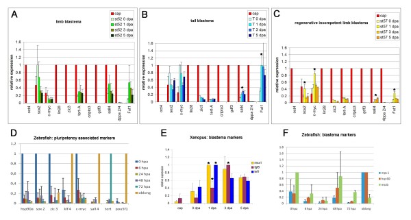
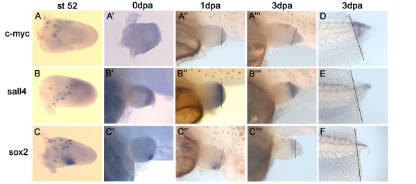
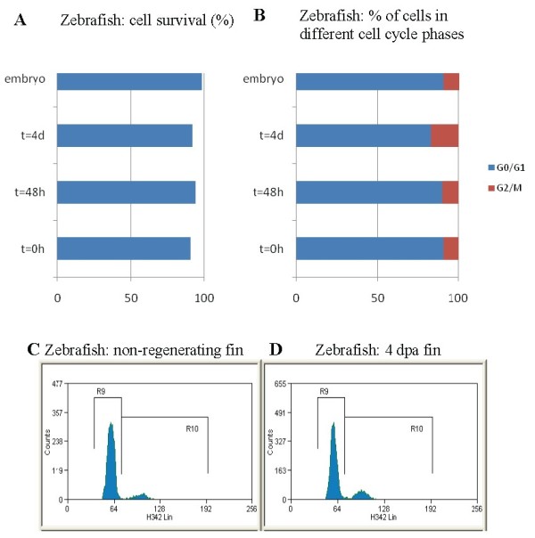
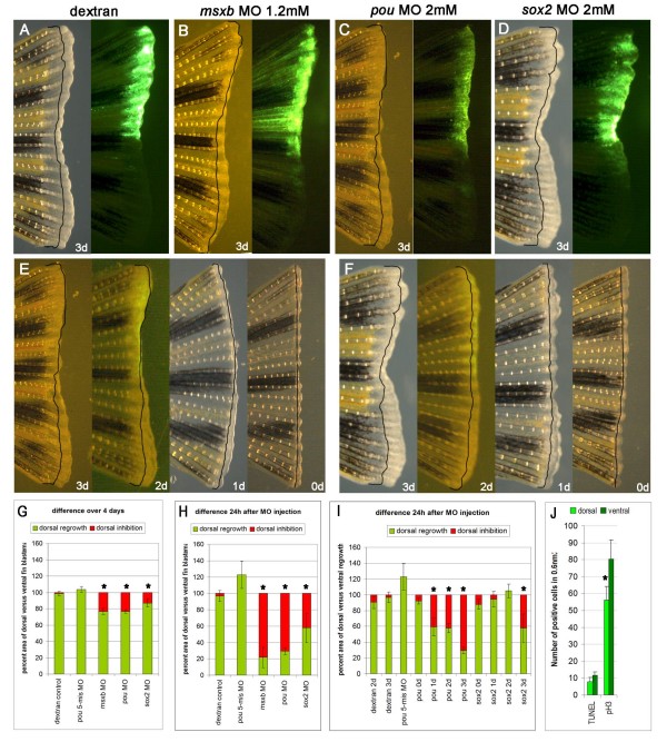
Results: We hypothesised that there are parallels between dedifferentiation or reprogramming of somatic cells to induced pluripotent stem cells and the natural process of dedifferentiation during epimorphic regeneration. We analysed expression levels of the most commonly used pluripotency associated factors in regenerating and non-regenerating tissue and compared them with levels in a pluripotent reference cell. We found that some of the pluripotency associated factors (oct4/pou5f1, sox2, c-myc, klf4, tert, sall4, zic3, dppa2/4 and fut1, a homologue of ssea1) were expressed before and during regeneration and that at least two of these factors (oct4, sox2) were also required for normal fin regeneration in the zebrafish. However these factors were not upregulated during regeneration as would be expected if blastema cells acquired pluripotency.
Conclusions: By comparing cells from the regeneration blastema with embryonic pluripotent reference cells we found that induced pluripotent stem and blastema cells do not share pluripotency. However, during blastema formation some of the key reprogramming factors are both expressed and are also required for regeneration to take place. We therefore propose a link between partially reprogrammed induced pluripotent stem cells and the half way state of blastema cells and suggest that a common mechanism might be regulating these two processes.
Figures
Similar articles
-
Dedifferentiation and the role of sall4 in reprogramming and patterning during amphibian limb regeneration.Dev Dyn. 2011 May;240(5):979-89. doi: 10.1002/dvdy.22554. Epub 2011 Feb 8. Dev Dyn. 2011. PMID: 21305648 Review.
-
Enhanced human somatic cell reprogramming efficiency by fusion of the MYC transactivation domain and OCT4.Stem Cell Res. 2017 Dec;25:88-97. doi: 10.1016/j.scr.2017.10.014. Epub 2017 Oct 26. Stem Cell Res. 2017. PMID: 29125994
-
An integrated systems biology approach identifies positive cofactor 4 as a factor that increases reprogramming efficiency.Nucleic Acids Res. 2016 Feb 18;44(3):1203-15. doi: 10.1093/nar/gkv1468. Epub 2016 Jan 5. Nucleic Acids Res. 2016. PMID: 26740582 Free PMC article.
-
A Versatile In Vivo System to Study Myc in Cell Reprogramming.Methods Mol Biol. 2021;2318:267-279. doi: 10.1007/978-1-0716-1476-1_14. Methods Mol Biol. 2021. PMID: 34019296
-
[The dual role of OCT4].Med Sci (Paris). 2010 Apr;26(4):411-6. doi: 10.1051/medsci/2010264411. Med Sci (Paris). 2010. PMID: 20412747 Review. French.
Cited by
-
Regenerative neurogenesis: the integration of developmental, physiological and immune signals.Development. 2022 Apr 15;149(8):dev199907. doi: 10.1242/dev.199907. Epub 2022 May 3. Development. 2022. PMID: 35502778 Free PMC article.
-
Transdifferentiation from cornea to lens in Xenopus laevis depends on BMP signalling and involves upregulation of Wnt signalling.BMC Dev Biol. 2011 Sep 6;11:54. doi: 10.1186/1471-213X-11-54. BMC Dev Biol. 2011. PMID: 21896182 Free PMC article.
-
Oct4 Methylation-Mediated Silencing As an Epigenetic Barrier Preventing Müller Glia Dedifferentiation in a Murine Model of Retinal Injury.Front Neurosci. 2016 Nov 15;10:523. doi: 10.3389/fnins.2016.00523. eCollection 2016. Front Neurosci. 2016. PMID: 27895551 Free PMC article.
-
A case of cellular alchemy: lineage reprogramming and its potential in regenerative medicine.J Mol Cell Biol. 2012 Aug;4(4):190-6. doi: 10.1093/jmcb/mjs005. Epub 2012 Feb 27. J Mol Cell Biol. 2012. PMID: 22371436 Free PMC article. Review.
-
Mechanisms of urodele limb regeneration.Regeneration (Oxf). 2017 Dec 26;4(4):159-200. doi: 10.1002/reg2.92. eCollection 2017 Aug. Regeneration (Oxf). 2017. PMID: 29299322 Free PMC article. Review.
References
-
- Satoh A, Bryant SV, Gardiner DM. Regulation of dermal fibroblast dedifferentiation and redifferentiation during wound healing and limb regeneration in the Axolotl. Development, Growth & Differentiation. 2008;50:743–754. - PubMed
Publication types
MeSH terms
Substances
LinkOut - more resources
Full Text Sources
Other Literature Sources
Molecular Biology Databases
