. 2010 Jan 20:9:11.
doi: 10.1186/1476-4598-9-11.

MUC16 provides immune protection by inhibiting synapse formation between NK and ovarian tumor cells

Affiliations

MUC16 provides immune protection by inhibiting synapse formation between NK and ovarian tumor cells

Jennifer A A Gubbels et al. Mol Cancer. .

Abstract

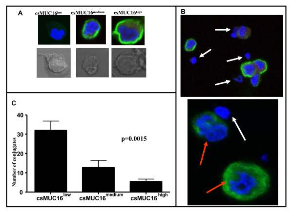
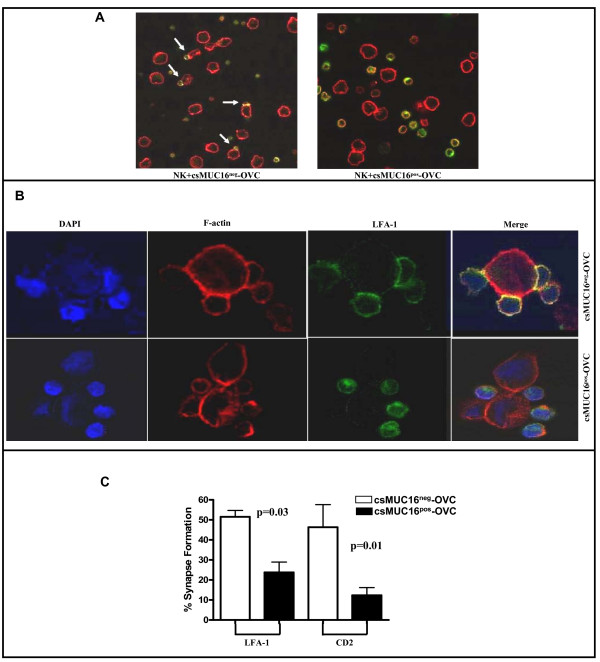
Background: Cancer cells utilize a variety of mechanisms to evade immune detection and attack. Effective immune detection largely relies on the formation of an immune synapse which requires close contact between immune cells and their targets. Here, we show that MUC16, a heavily glycosylated 3-5 million Da mucin expressed on the surface of ovarian tumor cells, inhibits the formation of immune synapses between NK cells and ovarian tumor targets. Our results indicate that MUC16-mediated inhibition of immune synapse formation is an effective mechanism employed by ovarian tumors to evade immune recognition.

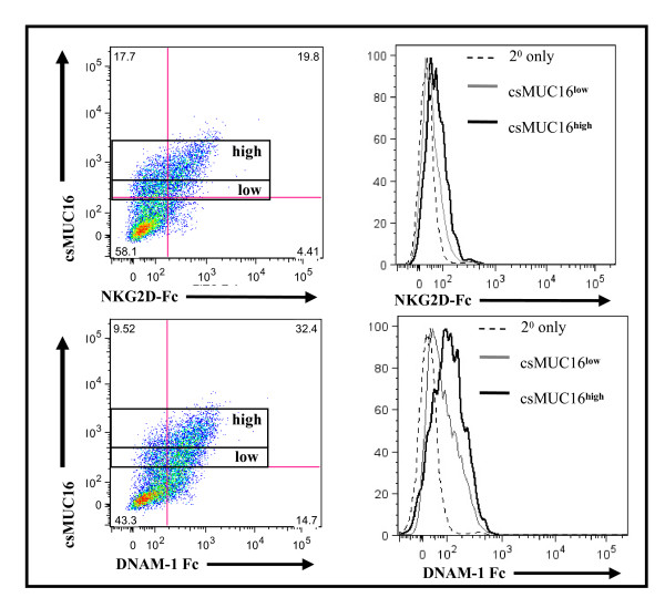
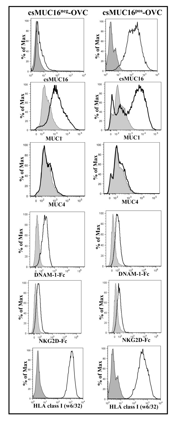
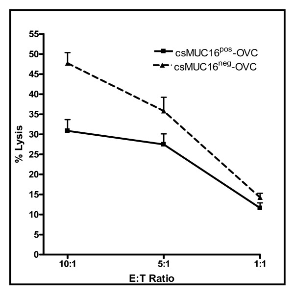
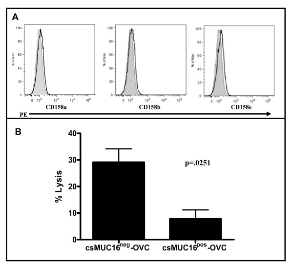
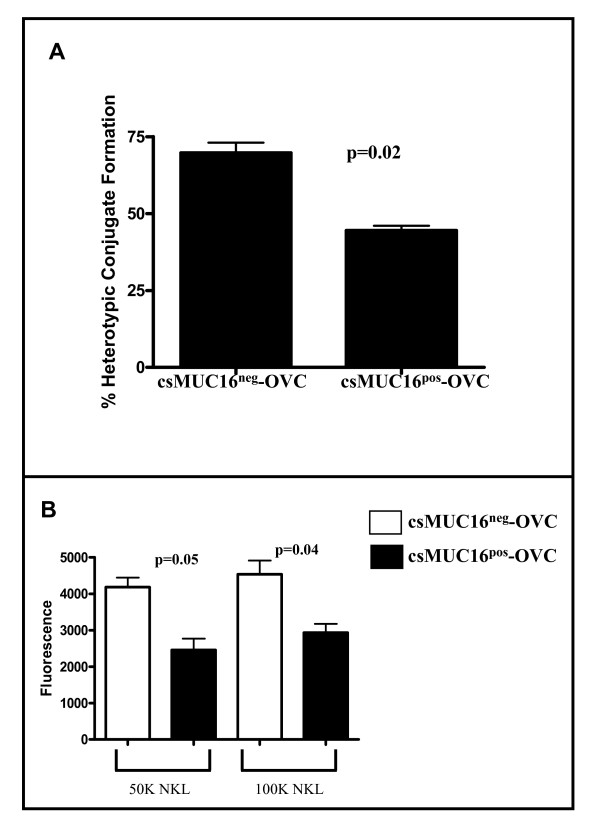
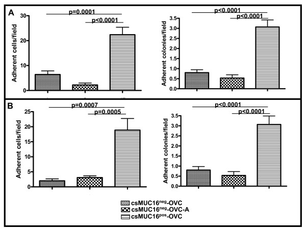
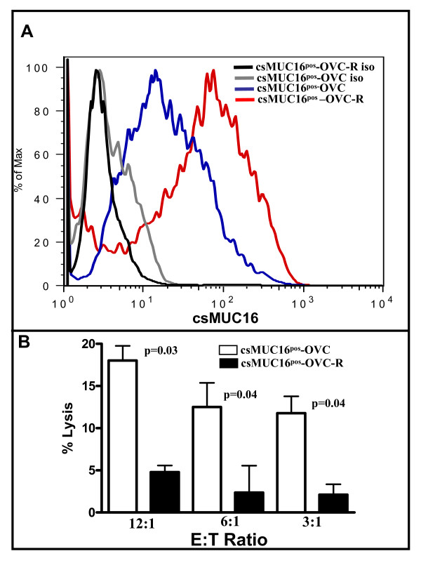
Results: Expression of low levels of MUC16 strongly correlated with an increased number of conjugates and activating immune synapses between ovarian tumor cells and primary naïve NK cells. MUC16-knockdown ovarian tumor cells were more susceptible to lysis by primary NK cells than MUC16 expressing controls. This increased lysis was not due to differences in the expression levels of the ligands for the activating receptors DNAM-1 and NKG2D. The NK cell leukemia cell line (NKL), which does not express KIRs but are positive for DNAM-1 and NKG2D, also conjugated and lysed MUC16-knockdown cells more efficiently than MUC16 expressing controls. Tumor cells that survived the NKL challenge expressed higher levels of MUC16 indicating selective lysis of MUC16(low) targets. The higher csMUC16 levels on the NKL resistant tumor cells correlated with more protection from lysis as compared to target cells that were never exposed to the effectors.

Conclusion: MUC16, a carrier of the tumor marker CA125, has previously been shown to facilitate ovarian tumor metastasis and inhibits NK cell mediated lysis of tumor targets. Our data now demonstrates that MUC16 expressing ovarian cancer cells are protected from recognition by NK cells. The immune protection provided by MUC16 may lead to selective survival of ovarian cancer cells that are more efficient in metastasizing within the peritoneal cavity and also at overcoming anti-tumor innate immune responses.


Figures

Figure 1
Figure 1
Selective targeting of csMUC16low OVCAR-3 cells by NK cells. A, Upper panels show the classification of csMUC16low, medium, and high OVCAR-3 cells. This classification was used to determine NK cell conjugation with different subsets of OVCAR-3 cells. The lower panel shows the corresponding bright field image of the cell in the upper panel. B, The upper panel is a 60× view of NK cells (white arrows) forming conjugates with csMUC16low cells. NK cells surrounding csMUC16high cells are not touching the tumor cell. The lower panel is a digital zoom image demonstrating selective conjugation of NK cells (white arrow) with csMUC16low and not with csMUC16high OVCAR-3 cells (red arrows). C, Conjugates of NK cells with low, medium and high csMUC16 expressing cells were quantified by averaging the number of conjugates observed per 50 fields. Data shown is mean of four independent experiments performed with NK cells from separate donors. A one-way ANOVA was performed and the resulting p value = .0015.
Figure 2
Figure 2
DNAM-1 and NKG2D ligand expression is comparable on csMUC16high expressing OVCAR-3 cells. OVCAR-3 cells were stained for csMUC16 expression, and then NKG2D-Fc or DNAM-1-Fc and an appropriate secondary antibody were added. Cells were initially gated on live single events of tumor cells that were csMUC16low or csMUC16high. The binding of NKG2D-Fc or DNAM-1 to gated csMUC16low (gray line) and csMUC16high (black line) is shown in histogram form. Data is representative of three independent experiments.
Figure 3
Figure 3
Expression of mucins and NK cell activating and inhibitory molecules on csMUC16neg-OVC and csMUC16pos-OVC. MUC1 expression on both sublines is similar and MUC4 expression is absent. Expression of DNAM-1 and NKG2D ligands on the sublines was determined by staining with Fc chimeras (15 μg/mL) of these two receptors followed by labeling with fluorophore conjugated anti-Fc secondary antibody. HLA class I expression on the sublines was determined by labeling with FITC conjugated w6/32. Histograms of live single events are shown in all plots. Data is representative of three independent experiments. Shaded peaks show control cells incubated with only the fluorescently-tagged secondary antibodies. Un-shaded histograms are for cells incubated with the primary antibodies or Fc chimeras and the fluorescently labeled secondary antibodies.
Figure 4
Figure 4
csMUC16 protects ovarian cancer cells from lysis by NK cells. NK cells from four healthy donors were isolated and 51Cr release lysis assays at 10:1, 5:1, and 1:1 effector:target ratios were conducted using csMUC16neg-OVC and csMUC16pos-OVC cells. Chromium release from the targets was determined after 4 h incubation under standard tissue culture conditions. Percent lysis was calculated by determining the spontaneous and maximum release of radioactivity from each cell line.
Figure 5
Figure 5
NKLs lack KIRs and exhibit reduced ability to lyse csMUC16pos-OVC. A Expression of KIR on NKL cells was determined by flow cytometry. B. Flow cytometry based cell lysis assays (24:1 effector:target ratio) were conducted to demonstrate that NKL cells lyse csMUC16neg-OVC cells to a greater extent than the csMUC16pos-OVC cells. Mean, standard deviation and p-value of three independent measurements is shown.
Figure 6
Figure 6
csMUC16neg-OVC form more conjugates with NK and NKL cells than csMUC16pos-OVC. A, NK cells from healthy donors were labeled with CellTracker Green and mixed with CellTracker Blue labeled csMUC16neg-OVC or csMUC16pos-OVC cells. Conjugation between the tumor cells and NK cells was determined by flow cytometry. Mean values of NK cell-tumor cell conjugation obtained from experiments conducted with three healthy donors are shown. B. csMUC16neg-OVC or csMUC16pos-OVC were plated in a 96-well plate and allowed to grow to 70-80% confluence. Calcein dyed NKLs were added to the plate. After incubation, non-adherent cells were removed by washing and the NKL cells bound to tumor targets were quantified using a fluorescence plate reader. Data for two experiments was combined for a total of 12 replicates.
Figure 7
Figure 7
csMUC16 inhibits NK activating synapse formation. A. NK cells from healthy donors were mixed with csMUC16neg-OVC and csMUC16pos-OVC cells and confocal microscopy was conducted using 10× magnification. White arrows indicate NK cells bound to tumor cells and some forming activating immune synapses. B. Immune synapses between the NK cells and csMUC16neg-OVC at 40× magnification are shown. NK cells are present in the vicinity of csMUC16pos-OVC cells but form significantly less conjugates and immune synapses. C. Immune synapses between NK cells from three healthy donors and the csMUC16neg-OVC or the csMUC16pos-OVC cells were quantified by counting conjugates that showed polarization of LFA-1 and CD2 divided by the total number of conjugates (defined as two cells in contact).
Figure 8
Figure 8
csMUC16 shields ovarian cancer cells from NKL mediated lysis. A. NKL cells were added to confluent cultures of csMUC16neg-OVC or csMUC16pos-OVC cells at a 1:1 effector:target ratio. After incubation for A, 24 h and B, 48 h the number of adherent colonies and adherent cells surviving the NKL challenge were counted by microscopic examination of five random fields. This experiment was repeated in triplicate and mean values (n = 15 fields) and standard deviation are shown.
Figure 9
Figure 9
Tumor cells surviving NKL challenge express higher levels of csMUC16. A. csMUC16pos-OVC cells not treated with NKL or those surviving NKL treatment (csMUC16pos-OVC-R) were analyzed for csMUC16 expression by flow cytometry. Data is representative of three independent experiments. B, csMUC16pos-OVC and csMUC16pos-OVC-R were labeled with 51Cr. Cytotoxity assays were performed by using NKL cells as effectors. After 4 h co-incubation, released radioactivity was measured. Each bar is mean of four independent assays.

Similar articles

Cited by

References

    1. Zhang L, Conejo-Garcia JR, Katsaros D, Gimotty PA, Massobrio M, Regnani G, Makrigiannakis A, Gray H, Schlienger K, Liebman MN, Rubin SC, Coukos G. Intratumoral T cells, recurrence, and survival in epithelial ovarian cancer. N Engl J Med. 2003;348:203–213. doi: 10.1056/NEJMoa020177. - DOI - PubMed
    1. Carlsten M, Bjorkstrom NK, Norell H, Bryceson Y, van Hall T, Baumann BC, Hanson M, Schedvins K, Kiessling R, Ljunggren HG, Malmberg KJ. DNAX accessory molecule-1 mediated recognition of freshly isolated ovarian carcinoma by resting natural killer cells. Cancer Res. 2007;67:1317–1325. doi: 10.1158/0008-5472.CAN-06-2264. - DOI - PubMed
    1. Carlsten M, Malmberg KJ, Ljunggren HG. Natural killer cell-mediated lysis of freshly isolated human tumor cells. Int J Cancer. 2009;124:757–762. doi: 10.1002/ijc.24082. - DOI - PubMed
    1. Taylor DD, Gercel-Taylor C, Lyons KS, Stanson J, Whiteside TL. T-cell apoptosis and suppression of T-cell receptor/CD3-zeta by Fas ligand-containing membrane vesicles shed from ovarian tumors. Clin Cancer Res. 2003;9:5113–5119. - PubMed
    1. Lai P, Rabinowich H, Crowley-Nowick PA, Bell MC, Mantovani G, Whiteside TL. Alterations in expression and function of signal-transducing proteins in tumor-associated T and natural killer cells in patients with ovarian carcinoma. Clin Cancer Res. 1996;2:161–173. - PubMed

Publication types

MeSH terms

Substances