. 2010;19(4):487-503.
doi: 10.3727/096368909X486057. Epub 2010 Jan 20.

Targeted disruption of outer limiting membrane junctional proteins (Crb1 and ZO-1) increases integration of transplanted photoreceptor precursors into the adult wild-type and degenerating retina

Affiliations

Targeted disruption of outer limiting membrane junctional proteins (Crb1 and ZO-1) increases integration of transplanted photoreceptor precursors into the adult wild-type and degenerating retina

R A Pearson et al. Cell Transplant. 2010.

Abstract

Diseases culminating in photoreceptor loss are a major cause of untreatable blindness. Transplantation of rod photoreceptors is feasible, provided donor cells are at an appropriate stage of development when transplanted. Nevertheless, the proportion of cells that integrate into the recipient outer nuclear layer (ONL) is low. The outer limiting membrane (OLM), formed by adherens junctions between Müller glia and photoreceptors, may impede transplanted cells from migrating into the recipient ONL. Adaptor proteins such as Crumbs homologue 1 (Crb1) and zona occludins (ZO-1) are essential for localization of the OLM adherens junctions. We investigated whether targeted disruption of these proteins enhances donor cell integration. Transplantation of rod precursors in wild-type mice achieved 949 +/- 141 integrated cells. By contrast, integration is significantly higher when rod precursors are transplanted into Crb1(rd8/rd8) mice, a model of retinitis pigmentosa and Lebers congenital amaurosis that lacks functional CRB1 protein and displays disruption of the OLM (7,819 +/- 1,297; maximum 15,721 cells). We next used small interfering (si)RNA to transiently reduce the expression of ZO-1 and generate a reversible disruption of the OLM. ZO-1 knockdown resulted in similar, significantly improved, integration of transplanted cells in wild-type mice (7,037 +/- 1,293; maximum 11,965 cells). Finally, as the OLM remains largely intact in many retinal disorders, we tested whether transient ZO-1 knockdown increased integration in a model of retinitis pigmentosa, the rho(-/-) mouse; donor cell integration was significantly increased from 313 +/- 58 cells without treatment to 919 +/- 198 cells after ZO-1 knockdown. This study shows that targeted disruption of OLM junctional proteins enhances integration in the wild-type and degenerating retina and may be a useful approach for developing photoreceptor transplantation strategies.


Figures
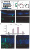
Figure 1
Figure 1
Integration of photoreceptors transplanted into the Crb1rd8/rd8 retina is higher than in wild-type controls. (A) Left, schematic showing the adherens junction complexes (red) between Müller glia terminal processes and photoreceptor inner segments, and right, some of the proteins involved in adherens junction formation and localization [adapted from (28)]. (B) Schematic of a subretinal injection. (C) Confocal projection montage showing integration into the 6-week-old Crb1rd8/rd8 mouse. Some cells remain in the subretinal space at 3 weeks posttransplantation but large numbers migrate into the ONL. (D) Histogram showing the number of integrated photoreceptors found at 3 weeks posttransplantation into 3-, 6-, or 12-week-old Crb1rd8/rd8 recipients, compared with 6-week-old wild-type recipients. Error bars: ±SEM. p-values: ANOVA with Bonferonni's correction for multiple comparisons. (E, F) Integrated cells were frequently observed in large clusters of 10–30 cells in the Crb1rd8/rd8 mouse (E). This contrasts integration into the wild-type retina, which is usually sporadic with single cells, or small clusters of 2–5 cells (F). Scale bars: 50 μm.
Figure 2
Figure 2
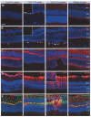
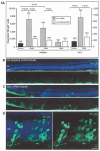
The abnormal OLM in the Crb1rd8/rd8 mouse leads to a progressive disorganization of the ONL and increasing Müller glial cell activation with age. (A–E) Immunohistochemistry showing confocal projection images of staining for proteins associated with adherens junctions, Crb1 (A) and ZO-1 (B), the cell adhesion marker, pancadherin (C), glial fibrilliary acidic protein (GFAP) (D, E), which labels activated astrocytes and glia and vimentin (E), which labels Müller glia regardless of activation (37). Staining is shown in 3-, 6-, and 12-week-old Crb1rd8/rd8 mice and 6-week-old wild-type mice. Insets in (A) and (B), single confocal sections from 6-week-old Crb1rd8/rd8 and wild-type mice showing the fragmented staining pattern (arrows) of ZO-1 and Crb1 in the Crb1rd8/rd8 mouse. Inset in (D), GFAP staining shows GFAP-positive fibers extending along the edge of the ONL at 12 weeks. GFAP staining increased markedly between 6 and 12 weeks. All images shown are taken from the superior retina. Scale bars: 50 μm and 10μm (insets).
Figure 3
Figure 3
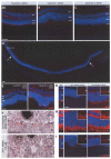
ZO-1 siRNA knocks down ZO-1 expression in the OLM. (A–C) Confocal images showing ZO-1 staining of the OLM following subretinal injection of 10 μM (A), 15 μM (B), or 20 μM (C). (D) Confocal montage showing extent of ZO-1 knockdown following application of 20 μM siRNA. Limits are marked by arrows. Significant OLM disruption was seen following application of the 15 μM dose, and knockdown was extensive with the 20 μM dose. (E) Apoptosis counts at 48 h postinjection in eyes receiving 20 or 15 μM ZO-1 siRNA, and over time in those receiving 15 μM siRNA, compared with nontargeting controls. (F) Electron micrographs showing disruption of the OLM 48 h after application of 15 μM ZO-1 siRNA or nontargeting control siRNA. Adherens junctions are highlighted by arrows. (G–I) Confocal images showing immunostaining for crb1 (G), pancadherin (H), and β-catenin (I), 48 h after application of 15 μM ZO-1 siRNA or nontargeting control siRNA. Wild-type sections are also included for comparison (left). Scale bars: 50 μm and 5 μm (F).
Figure 4
Figure 4
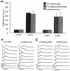
Retinal function is not impaired by ZO-1 siRNA administration. (A) Histogram showing average a- and b-wave amplitudes at a flash intensity of 0.1 cd s m−1. (B, C) ERG intensity series at 8 weeks following subretinal injection of either 15 μM ZO-1 siRNA (B, left) or 15 μM nontargeting control siRNA (C, left), compared with uninjected contralateral control eyes (A, right and B, respectively). ERG traces shown in each chart were recorded at intensities of 0.001, 0.01, 0.1, 1, 3, 5, and 10 cd s m−1. Error bars: ±SEM.
Figure 5
Figure 5
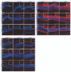
Time course of OLM disruption following subretinal injection of ZO-1 siRNA. Confocal images showing staining for ZO-1 (A–C), pancadherin (D–F), and GFAP (G–I) at 48 and 72 h and 1 week postinjection of 15 μM ZO-1 siRNA (A, D, G), 15 μM nontargeting control siRNA (B, E, H), or vehicle (C, F, I). Scale bar: 50 μm. Insets show high-power images of single confocal sections of ZO-1 or pancadherin immunolabeling, where appropriate. Arrowheads depict level of OLM. Scale bar: 10 μm.
Figure 6
Figure 6
Enhanced integration of transplanted photoreceptors following ZO-1 siRNA treatment. (A) Histogram showing the number of integrated rod photoreceptors found at 3 weeks posttransplantation of unsorted P4 Nrl.gfp+/+ donor cells into either 6-week-old wild-type recipients or 4-week-old rho−/− recipients, pretreated with ZO-1 siRNA (gray bars), compared with contralateral control eyes treated with equivalent concentrations of nontargeting control siRNA (white bars), and standard wild-type or rho−/− controls (no preinjection; black bars). Error bars: ±SEM. p-values: regular type = ANOVA with Bonferonni's correction for multiple comparisons; italic type = paired t-test. (B, C) Confocal projection montage showing integration of unsorted P4 Nrl.gfp+/+ donor cells into a 6-week-old wild-type mouse treated with nontargeting control siRNA (B) in one eye and ZO-1 siRNA in the other (C). (D) Integrated cells were frequently observed in large clusters of 10–30 cells in the ZO-1 siRNA-treated eyes.
Figure 7
Figure 7
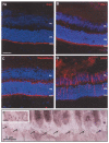
The OLM is intact in the 4-week-old rho−/− mouse. (A–D) Confocal images showing staining for ZO-1, Crb-1, pancadherin, and GFAP in the 1-month-old rho−/− mouse. Scale bar: 50 μm. (E) Electron micrograph of the OLM in the 3-month-old rho−/− mouse. Adherens junctions are highlighted by arrows. Scale bars: 5 μm and 1 μm (inset).

References

    1. Aijaz S, Balda MS, Matter K. Tight junctions: Molecular architecture and function. Int. Rev. Cytol. 2006;248:261–298. - PubMed
    1. Akimoto M, Cheng H, Zhu D, Brzezinski JA, Filippova E, Oh ECT, Jing Y, Linares J-L, Brooks M, Zareparsi S, Mears AJ, Hero A, Glaser T, Swaroop A. Targeting of green fluorescent protein to new-born rods by Nrl promoter and temporal expression profiling of flow-soreted photoreceptors. Proc. Natl. Acad. Sci. USA. 2006;103(10):3890–3895. - PMC - PubMed
    1. Bartsch U, Oriyakhel W, Kenna PF, Linke S, Richard G, Petrowitz B, Humphries P, Farrar GJ, Ader M. Retinal cells integrate into the outer nuclear layer and differentiate into mature photoreceptors after subretinal transplantation into adult mice. Exp. Eye Res. 2008;86:691–700. - PubMed
    1. Campbell M, Humphries M, Kenna P, Humphries P, Brankin B. Altered expression and interaction of adherens junction proteins in the developing OLM of the Rho(−/−) mouse. Exp. Eye Res. 2007;85:714–720. - PubMed
    1. Campbell M, Humphries M, Kennan A, Kenna P, Humphries P, Brankin B. Aberrant retinal tight junction and adherens junction protein expression in an animal model of autosomal dominant Retinitis pigmentosa: The Rho(−/−) mouse. Exp. Eye Res. 2006;83:484–492. - PubMed

Publication types

MeSH terms

LinkOut - more resources