. 2011 Jun 15;168(2):325-33.e1.
doi: 10.1016/j.jss.2009.09.030. Epub 2009 Oct 9.

A novel role for NKT cells in cutaneous wound repair

Affiliations

A novel role for NKT cells in cutaneous wound repair

David F Schneider et al. J Surg Res. .

Abstract

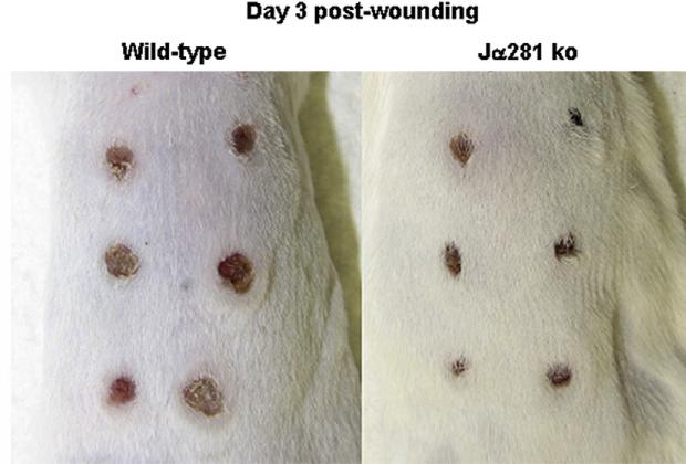
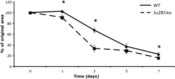
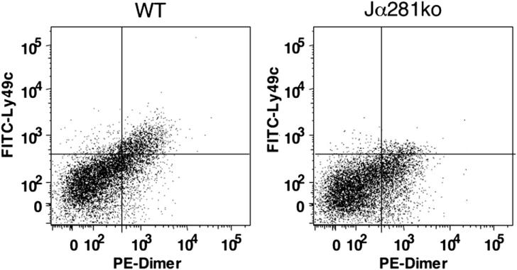
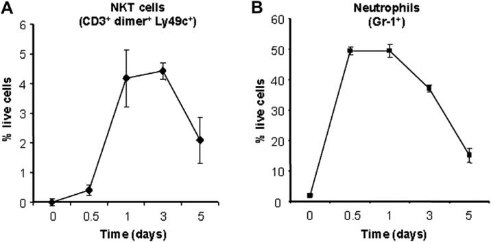
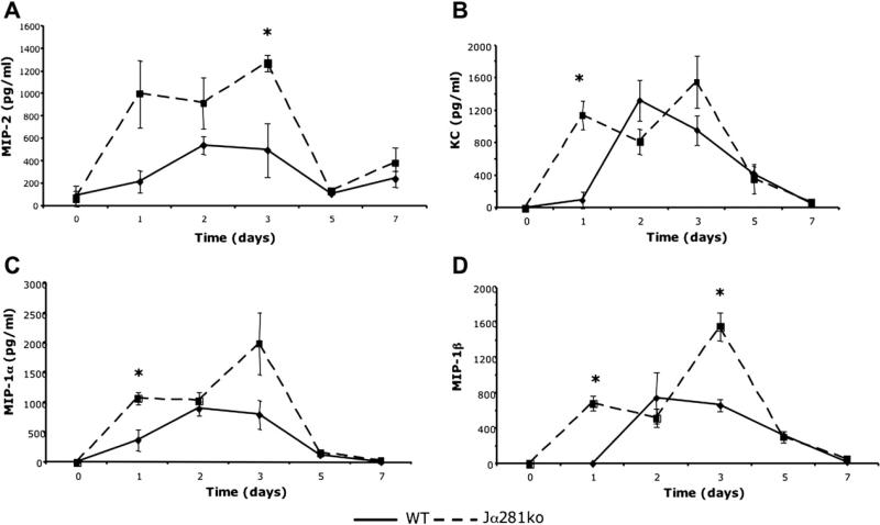
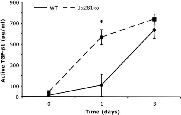
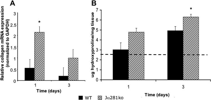
Here, we report the novel observation that natural killer T (NKT) cells contribute to the cutaneous wound repair process. Using an excisional wound model in wild-type versus NKT cell-deficient mice, this report shows that when NKT cells are absent, initial wound closure is markedly accelerated. We report here for the first time that NKT cells are a significant constituent of early wound inflammation and that they regulate the local production of a key subset of neutrophil and monocyte/macrophage chemokines, as well as TGF-β1 content and collagen deposition. Together, our findings support the concept that NKT cells regulate the early inflammatory and fibroproliferative phases of nonpathologic healing wounds, positioning the NKT cell as an attractive potential therapeutic target for modulation of impaired wound healing.


Figures

FIG. 1
FIG. 1
Comparison of excisional wound closure in wild type versus NKT cell deficient mice. Wild type BALB/c and BALB/c-Jα281ko mice were each given six 3 mm dorsal punch excisional wounds. Three days later, wounds were photographed; n = 3–6 per group. Similar results were obtained from two separate experiments for WT versus Jα281ko and two separate experiments for WT versus CD1dko. (Color version of figure is available online.)
FIG. 2
FIG. 2
Wound closure wild-type versus NKT cell-deficient Jα281ko mice. Wild type BALB/c and BALB/c-Jα281ko mice were each given six 3 mm dorsal punch excisional punch wounds. At d 1, 3, 5, and 7 post-wounding, pelts were removed and the individual wounds photographed. The percent open wound area was calculated from the number of pixels in the open wound area. *P < 0.05, n = 6 per group. Experiments were done three times.
FIG. 3
FIG. 3
Day 1 wound NKT cell infiltrates in wild type versus Jα281ko mice. Day 1 wound dermal cell suspensions were immunostained with APC CD3, PE IgG:CD1d dimeric fusion protein, and FITC Ly49c and analyzed by flow cytometry. After gating on the CD3+ population, NKT cells were identified as those positive for Ly49c and intermediate staining of CD1d dimer. One representative sample per group is shown. Data are shown as percentage of live cells that are CD3 + IgG:CD1d dimeric fusion protein+ Ly49c+. The cumulative data from this experiment are shown in Fig. 4.
FIG. 4
FIG. 4
Wound NKT cell versus neutrophil content over time. Wild type BALB/c mice were given 3 mm excisional punch wounds. At 12 h and 1, 3, and 5 d post-wounding, animals were sacrificed, their wounds collected, and dermal cell suspensions created. NKT cells were identified by flow cytometric analysis of dermal cell suspensions as those staining positively for CD3, Ly49c, and glycolipid-loaded IgG:CD1d dimeric fusion protein (A). Neutrophils were identified via flow cytometry for Gr-1+ cells (B). Dermal cell suspensions created from the dorsal skin of uninjured animals were also analyzed (d 0). Data are presented as the percentage of live cells ± SEM; n = 4 animals per group.
FIG. 5
FIG. 5
Wound chemokine content in WT versus Jα281ko mice. WT BALB/c and BALB/c-Jα281ko mice were given 3 mm excisional punch wounds and wound biopsies were collected 1, 2, 3, 5, and 7 d later. Skin biopsies from uninjured animals were used as time 0 controls. Wounds were homogenized in protease inhibitor cocktail and chemokine content was determined by ELISA. Data are represented as mean pg chemokine per mL of homogenate; n = 3–4 mice per group; *P < 0.05.
FIG. 6
FIG. 6
Wound TGF-β1 content in WT versus Jα281ko mice. WT BALB/c and BALB/c-Jα281ko mice were given 3 mm excisional punch wounds, and wound biopsies were collected 1 and 3 d later. Skin biopsies from uninjured animals were used as time 0 controls. Wounds were homogenized in protease inhibitor cocktail. The active fraction of TGF-β1 was determined by ELISA. Data are represented as mean pg TGF-β1 per mL of homogenate; n = 4 mice per group; *P < 0.05.
FIG. 7
FIG. 7
Wound α2(I)-procollagen gene expression and collagen content in WT versus Jα281ko mice. WT BALB/c and BALB/c-Jα281ko mice were given 3 mm punch wounds and at d 1 and 3, wound mRNA expression for α2(I)-procollagen and GAPDH was quantitated by real-time polymerase chain reaction (PCR). Relative procollagen mRNA expression was determined by normalization of each sample to GAPDH expression (A). Wounds from the same animals were hydrolyzed in 6 N HCL and subjected to the hydroxyproline assay to quantitate wound collagen content (B); n = 4 mice per group; *P < 0.05.

Similar articles

Cited by

References

    1. Joyce S. CD1d and natural T cells: How their properties jump-start the immune system. Cell Molec Life Sci. 2001;58:442. - PMC - PubMed
    1. Kawakami K, Yamamoto N, Kinjo Y, et al. Critical role of Vα14+ natural killer T cells in the innate phase of host protection against Streptococcus pneumoniae infection. Eur J Immunol. 2003;33:3322. - PubMed
    1. Nieuwenhuis EE, Matsumoto T, Exley MA, et al. CD1d-dependent macrophage-mediated clearance of Pseudomonas aeruginosa from lung. Nature Med. 2002;8:588. - PubMed
    1. Faunce DE, Stein-Streilein J. NKT cell-derived RANTES recruits APCs and CD8 + T cells to the spleen during the generation of regulatory T cells in tolerance. J Immunol. 2002;169:31. - PubMed
    1. Wilson SB, Kent SC, Patton KT, et al. Extreme Th1 bias of invariant Vα24 JαQ T cells in type 1 diabetes. Nature. 1998;1998(391):177. - PubMed

Publication types