Review
. 2010 Mar 2;9(3):286-302.
doi: 10.1016/j.dnarep.2009.12.006. Epub 2010 Jan 20.

Rad54, the motor of homologous recombination

Affiliations
Review

Rad54, the motor of homologous recombination

Alexander V Mazin et al. DNA Repair (Amst). .

Abstract

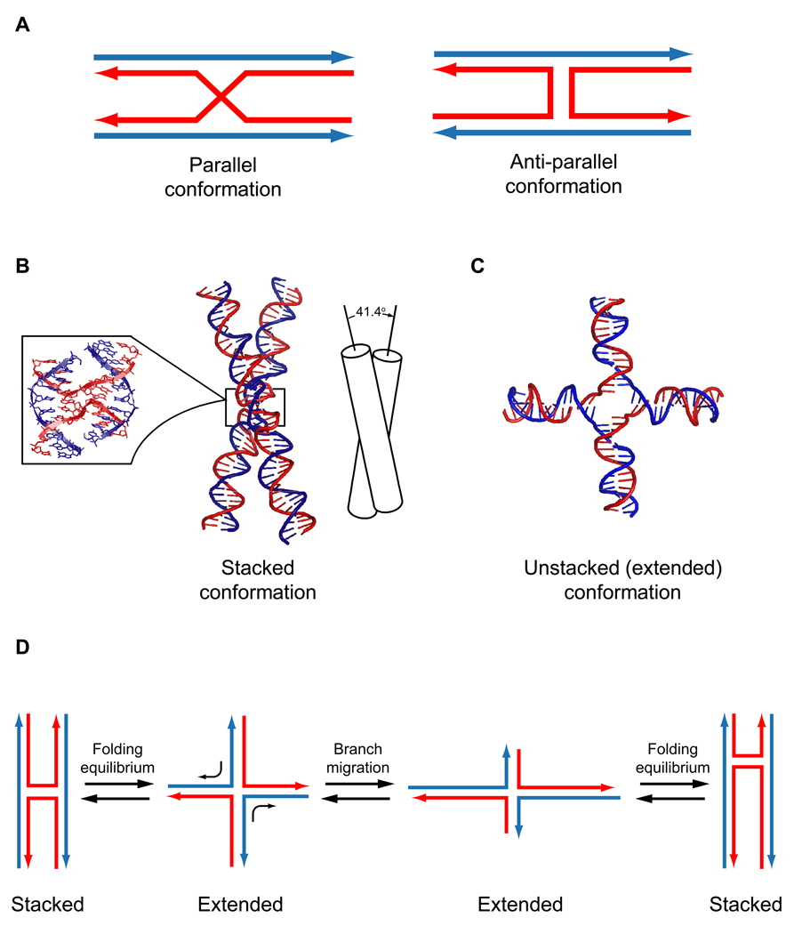
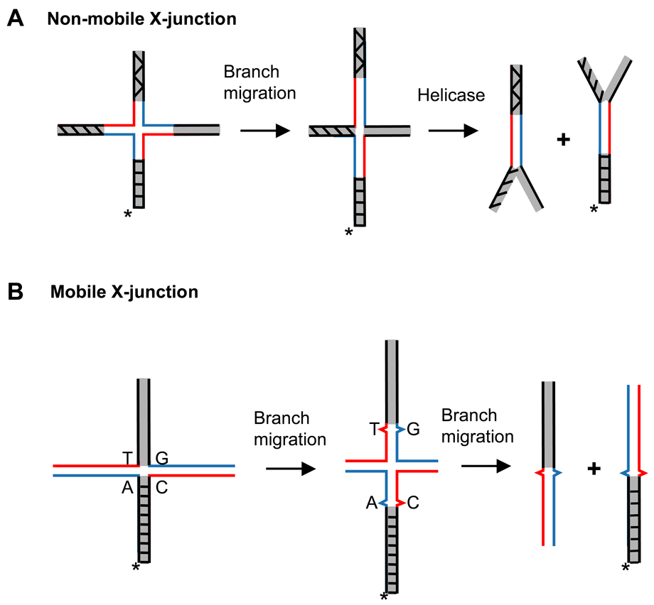
Homologous recombination (HR) performs crucial functions including DNA repair, segregation of homologous chromosomes, propagation of genetic diversity, and maintenance of telomeres. HR is responsible for the repair of DNA double-strand breaks and DNA interstrand cross-links. The process of HR is initiated at the site of DNA breaks and gaps and involves a search for homologous sequences promoted by Rad51 and auxiliary proteins followed by the subsequent invasion of broken DNA ends into the homologous duplex DNA that then serves as a template for repair. The invasion produces a cross-stranded structure, known as the Holliday junction. Here, we describe the properties of Rad54, an important and versatile HR protein that is evolutionarily conserved in eukaryotes. Rad54 is a motor protein that translocates along dsDNA and performs several important functions in HR. The current review focuses on the recently identified Rad54 activities which contribute to the late phase of HR, especially the branch migration of Holliday junctions.


Figures

Fig. 1
Fig. 1. DNA double strand break (DSB) repair by HR
Initial steps of HR (see explanations in the text) result in formation of Joint molecules (D-loops) that are further extended by DNA polymerase and processed afterward through the two major pathways, SDSA or DSBR. (A) Repair of DSB through the synthesis dependent strand annealing (SDSA) mechanism resuls in non-crossover recombinants. Dissociation of extended D-loop by branch migration is a crucial step of the SDSA mechanism. (B) The double-stranded break repair (DSBR) mechanism, which is more frequent during meiosis, results in crossover recombinants. DSB repair by HR generates Holliday junctions, which may branch migrate along the DNA axis. After second end capture, branch migration would either stabilize double D-loops by increasing the length of DNA heteroduplex or cause their dissociation channeling recombination intermediates through the double D-loop dissociation (DDD) mechanism into the SDSA pathway [132].
Fig. 2
Fig. 2. Structural domains of Rad54
(A) Schematic diagram highlighting the conserved SF2 motifs of Rad54. The RecA-like domains are shown in red with the seven “signature” motifs (along with the TxGx domain) displayed in blue. The single letter abbreviations above the motifs blocks emphasize the location of conserved amino acid residues, whose role in ATP hydrolysis is well-defined for SF2 family proteins. The NTD (the N-terminal domain) and the CTD (the C-terminal domain) are displayed in tan. (B) Schematic diagram highlighting the conserved motifs of Rad54 that define the Snf2 family of proteins; with the Snf blocks shown in green (designated from “A” to “N” by the order of identification), and the HD (helical domain) motifs in purple. The TxGx motif was first designated as the Snf2 family specific motif Snf-A [24], but was later reclassified as TxGx when this motif was found to be conserved throughout the SF2 family [88]. Motifs are shown in relative position to one another, but are not drawn to scale. (C) Sequence alignment of the N-terminal domain of Rad54 from Homo sapiens (Human), Mus musculus (Mouse), Xenopus tropicalis (Frog), Danio rerio (Zebrafish), Drosophila melanogaster (Fruit Fly), and Saccharomyces cerevisiae (Yeast). Residues that match the consensus sequence are highlighted in yellow, and the residues that are similar to the consensus are highlighted in blue. (D) The X-ray crystal structure of C-terminal domain of zebrafish Rad54 (PDB code: 1Z3I) [24]. This domain is specific to the Rad54 protein and contains a zinc-coordinating motif. The residues involved in zinc ion coordination are labeled and represented as sticks in the model.
Fig. 3
Fig. 3
Model of Rad54-induced supercoiling. Rad54 (represented as the blue shape; figure-eight represents the dual RecA-like domains, and the square represents the N-terminal domain) is shown bound to covalently closed circular plasmid DNA. Supercoils may accumulate on this type of DNA substrate if two (or more) active Rad54 complexes are bound to the same DNA molecule and move in opposing directions. As the Rad54 complexes translocate (direction of translocation is indicated with arrows), positive supercoils (+) would accumulate in front of complexes and negative supercoils (−) would accumulate behind.
Fig. 4
Fig. 4
Rationalizing the ability of Rad54 to both stabilize and disrupt the Rad51 filament. The Rad54 protein (blue shape) contains two distinct binding sites. The N-terminal domain of Rad54 (square) specifically interacts with Rad51 (green oval). This interaction is thought to stabilize individual Rad51 monomers, which adds to the overall stability of the filament. On the other hand, the dual RecA-like domains of Rad54 (figure eight) form the binding site for dsDNA. As Rad54 translocates on the dsDNA (direction of translocation is indicated with arrows), it will displace the Rad51 monomers, thereby disrupting the filament.
Fig. 5
Fig. 5. Schematic representation of the 4-stranded (A) and 3-stranded (B) branch migration
X-junctions and PX-junctions were used as substrates in these two reactions, respectively.
Fig. 6
Fig. 6. Conformational forms of the Holliday junction
(A) The parallel and antiparallel conformation of the Holliday junction. The antiparallel conformation differs from the parallel one by a 180° rotation of one of the exchanging dsDNA molecule around the junction. (B) Holliday junctions were crystallized in the stacked anti-parallel conformation [142]. For clarity, the insert (left) contains a close up view of the crossover point of the Holliday junction. Two stacked duplexes form a right-handed twist with an angle of 41.4° (right). (C) A Holliday junction in the unstacked (extended) conformation with four arms directed toward the corners of the square. (D) The scheme shows folding equilibrium between the stacked and extended conformations of Holliday junction, which depends on the concentration of divalent metal ions [138]. Branch migration occurs while the Holliday junction is in the extended state and stops when the molecule adopts the staked conformation.
Fig. 7
Fig. 7. Holliday junction substrates for branch migration proteins
(A) Non-movable or partially movable X-junctions require DNA helicase activity for their dissociation. These substrates have been previously used for analysis of branch migration activity of prokaryotic branch migration proteins, RuvAB and RecG, and for RecQ helicases, which combine branch migration with conventional DNA helicase activity. (B) Movable X-junctions (can branch migrate in only one direction) were designed for Rad54 that does not possess DNA helicase activity. Movable junctions contain a mismatch to block spontaneous branch migration. Shaded regions denote heterologous DNA terminal branches. 32P-label at the DNA 5’-end (indicated by the asterisk) is required for visualization of the products of branch migration after gel-electrophoresis in polyacrylamide gels.
Fig. 8
Fig. 8. The kinetics of branch migration by HsRad54 and E. coli RuvAB (Mazina and Mazin, unpublished results)
(A) Experimental scheme. (B) Joint molecules were produced by using either RecA or HsRad51 recombinases in 4-strand exchange reactions and then deproteinized, as described [29,200]. The joint molecules (0. 3 nM) made by RecA or HsRad51 were used for branch migration by RuvAB (140 nM RuvA and 360 nM RuvB) at 37 °C or HsRad54 (200 nM) at 30 °C, respectively. The reaction conditions were the same, as described previously [29,201] The DNA products were analyzed by electrophoresis in agarose gels and quantified using a Storm 840 Phosphor Imager. Data are the mean of three experiments. Error bars represent standard error of the mean. RuvAB was purchased from BioAcademia Inc., Japan.
Fig. 9
Fig. 9. HsRad54 promotes dissociation of joint molecules (D-loops)
HsRad54 promotes dissociation of native (non-deproteinized) single D-loops produced by HsRad51 (denoted by green circles) (A) or native double D-loops produced by RAD51 and RAD52 proteins (B). The green arrow marks formation of the Holliday junction during branch migration. The asterisk indicates 32P label.
Fig. 10
Fig. 10. Activities of Rad54
Rad54 possesses a unique set of biochemical activities that allow it to function at every stage along the HR pathway. (A) The dsDNA translocation activity (shown by the accumulation of supercoiling in DNA) is thought to be required for most, if not all other Rad54 activities. (B) The chromatin remodeling and protein displacement activities free DNA for processing by other proteins. (C) Rad54 stimulates the DNA strand exchange activity of Rad51 by forming a specific complex with this protein. (D) Through its branch migration activity, Rad54 processes HR intermediates. (E) Rad54 contributes to the resolution of these intermediates by stimulating the endonuclease activity of Mus81-Eme1.

Similar articles

Cited by

References

    1. Krogh BO, Symington LS. Recombination proteins in yeast. Annu Rev Genet. 2004;38:233–271. - PubMed
    1. San Filippo J, Sung P, Klein H. Mechanism of eukaryotic homologous recombination. Annu Rev Biochem. 2008;77:229–257. - PubMed
    1. Pâques F, Haber JE. Multiple pathways of recombination induced by double-strand breaks in Saccharomyces cerevisiae. Microbiology and Molecular Biology Reviews. 1999;63:349–404. - PMC - PubMed
    1. Neale MJ, Keeney S. Clarifying the mechanics of DNA strand exchange in meiotic recombination. Nature. 2006;442:153–158. - PMC - PubMed
    1. Bishop DK. Multiple mechanisms of meiotic recombination. Cell. 2006;127:1095–1097. - PubMed

Publication types