. 2010 May;38(9):3106-18.
doi: 10.1093/nar/gkp1216. Epub 2010 Jan 19.

Structural and mechanistic insights into Helicobacter pylori NikR activation

Affiliations

Structural and mechanistic insights into Helicobacter pylori NikR activation

C Bahlawane et al. Nucleic Acids Res. 2010 May.

Abstract

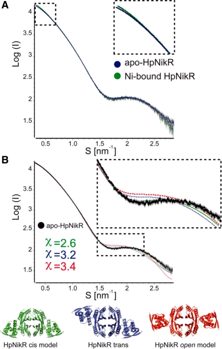
NikR is a transcriptional metalloregulator central in the mandatory response to acidity of Helicobacter pylori that controls the expression of numerous genes by binding to specific promoter regions. NikR/DNA interactions were proposed to rely on protein activation by Ni(II) binding to high-affinity (HA) and possibly secondary external (X) sites. We describe a biochemical characterization of HpNikR mutants that shows that the HA sites are essential but not sufficient for DNA binding, while the secondary external (X) sites and residues from the HpNikR dimer-dimer interface are important for DNA binding. We show that a second metal is necessary for HpNikR/DNA binding, but only to some promoters. Small-angle X-ray scattering shows that HpNikR adopts a defined conformation in solution, resembling the cis-conformation and suggests that nickel does not trigger large conformational changes in HpNikR. The crystal structures of selected mutants identify the effects of each mutation on HpNikR structure. This study unravels key structural features from which we derive a model for HpNikR activation where: (i) HA sites and an hydrogen bond network are required for DNA binding and (ii) metallation of a unique secondary external site (X) modulates HpNikR DNA binding to low-affinity promoters by disruption of a salt bridge.


Figures

Figure 1.
Figure 1.
Structural organization of Nickel bound HpNikR. (Left) Ribbon diagram of the overall Ni-HpNikR structure (14) in a view parallel to the crystallographic 2-fold axis. The tetramerization domain (TD) is coloured in black (chains A and C) and grey (chains B and D) and DNA-binding domains (DBD) in red. Nickel ions bound to high affinity (HA), intermediary (I) and external (X) sites are represented as spheres coloured in orange, yellow and green, respectively. (Right) Detailed view of the Ni(II)-binding sites HA and X and of the TD interface. Side chains of residues involved are represented in ball and stick. The residues mutated are indicated in the colour corresponding to the mutant: M1 (H99A, H101A, C107A), orange; M5 (H74G, H75G), green; M6 (C96S) brown; M7 (Q87F), violet and M10 (Q76A, R77A), pink. This colour-coding will be used in all the figures unless specified.
Figure 2.
Figure 2.
Ni(II) binding to NikR and M5, M6, M7 and M10 derivatives and kinetics of metal release for HpNikR and derivatives. (A) Comparison of the binding profiles of HpNikR, M5, M6, M7 and M10 determined by the changes in absorbance at 305 nm upon the addition of up to 3 equivalents of NiSO4. The presented data correspond to the difference in the absorption at 305 nm between the absorption spectra of the holo and apo forms for each protein. Data were collected at the equilibrium, 30 min after addition of NiSO4 to 200 µM of protein solution in HEPES 20 mM, pH 7.4. The results are homogenous (variation is <5%) and were reproduced at least three times with two different protein batches. (B) Comparison of the kinetics of Ni(II) release from HpNikR, M5, M6, M7 and M10. UV-visible absorption spectra were continuously measured for 1 h after EDTA addition. The changes in absorbance at 305 nm are presented as function of time after addition of 10 mM EDTA to a Ni(II)–protein complex with a 1 : 1 stoichiometry. Experiments were reproduced three times giving similar results.
Figure 3.
Figure 3.
DNA-binding properties of HpNikR and derivatives to PnixA. (A) Titration of the nixA affinity of HpNikR and mutants by EMSA with increasing protein concentrations from 1 to 40 nM against 450 pM PnixA. Binding buffer contained 100 µM NiSO4 while gel and running buffers contained 100 µM MnSO4. (B) Binding curves corresponding to the average fraction of bound PnixA related to the total labelled DNA were fitted using the Hill equation with an n-value of 1.5. Each plot is the average of four data sets. Variations were <5%.
Figure 4.
Figure 4.
DNA-binding properties of HpNikR to PnixA, PureA and PexbB in Ni-only and Ni/Mn conditions. The condition described in (A) corresponds to the one described by Zambelli et al. (13) with binding reactions performed in HEPES binding buffer [20 mM HEPES pH 7.85, 50 mM KCl, 0.01% Triton X-100, 0.1 mM DTT, 10% glycerol (v/v), 6 µg ml−1 sonicated sperm DNA, 100 µM NiSO4]. After a 15 min incubation with increasing HpNikR concentrations, complexes were separated on 5% acrylamide/bisacrylamide (19:1) gels, in MOPS/NaOAc running buffer (20 mM MOPS, 5 mM NaOAc pH 7.0), run at room temperature for 110 min at 170 V. EMSA conditions in (B) were Bis–Tris buffer and the binding conditions described in ‘Materials and Methods’ section of the manuscript (binding buffer with NiSO4 and running buffer with MnSO4).
Figure 5.
Figure 5.
Western blots of total lysates of H. pylori WT 26695, M5 and M6 in response to increasing NiCl2 concentrations. Ten micrograms of total proteins were extracted from cultures grown to the mid-log phase in the absence or presence of NiCl2 and loaded on polyacrylamide gels, transferred to nitrocellulose membranes and revealed with antibodies directed against UreA and NikR. Coomassie-stained gels were used as loading control and are presented in Supplementary Figure S3.
Figure 6.
Figure 6.
Small-angle X-ray diffraction (SAXS) of both apo and Ni-bound HpNikR. (A) Experimental scattering profiles from the apo (blue circles) and holo (green circles) HpNikR showing that both curves are almost equivalent. Dashed line square shows a close-up of the low-angle region of both experimental curves. (B) Experimental scattering profile from the apo-HpNikR (black spheres) compared with the theoretical curves calculated for HpNikR in closed cis- (green), closed trans- (blue) and open (red) conformations. The best fit is obtained with the closed cis-conformation as shown by the chi between experimental and theoretical curves obtained using Crysol.
Figure 7.
Figure 7.
Apo- and Ni-bound structures of M5 reveal an absence of X sites. (A) Structural comparison of apo-M5 (blue) with Ni-M5 structure (green). (B) Detailed comparison of Ni-M5 structure (green) with Ni1-HpNikR (grey). Ni-M5 structure was superimposed onto Ni1-HpNikR [r.m.s deviation of 0.98 Å (531 Cα)]. Nickel ions bound to HA, I and X sites are indicated as sphere and coloured as in Figure 1. Amino acids involved in nickel binding are shown as balls and stick and coloured by atom type (carbon as the ribbon, green; oxygen, red; nitrogen, blue; and sulphur, orange). Nickel coordination and hydrogen bonds are presented as full lines and green broken lines, respectively. Residues from M5 mutant are labelled in red.
Figure 8.
Figure 8.
Apo-M7 structure suggests a weakening of β3–β′3 interactions and the disruption of the hydrogen bond network. (A) Detailed comparison of apo-HpNikR (grey) with apo-M7 (violet) in a view perpendicular to the 2-fold crystallographic axis showing the modifications induced by Q87F mutation (F87 side chain is indicated in red) at the tetramerization interface. The M7 structure was superimposed onto apo-HpNikR TD [r.m.s deviation of 1.76 Å (308 Cα)]. Accommodation of the F87 residue is accompanied by the displacement of α3 and α4 helices and the unzipping of β3–β′3 interactions at each side of the TD. These modifications unlock the TD interface and are echoed to the DBDs that adopt a modified closed trans-conformation. (B). Detailed comparison of the conserved hydrogen bond network linking two nickel ions at the HA sites within each ACT-like pair observed in Ni-EcNikR (pdb code 2HZV) and Ni1-HpNikR structures.
Figure 9.
Figure 9.
X sites tune the DNA-binding properties of HpNikR through the stabilization of β2–α3 loop and R77 availability. (Top) Theoretical model of the Ni–HpNikR–DNA ternary complex based on Ni–EcNikR–DNA complex. (Bottom) Detailed view of a model of HpNikR in closed cis-conformation bound to DNA, displayed in ribbon representation with chains B/D and A/C of HpNikR coloured in red and blue, respectively. Nickel ions bound to HA and X sites are indicated as sphere and coloured in orange and green, respectively. Amino acids from the TD involved DNA contacts are shown as balls and stick and coloured by atom type (carbon depending on the chain, green; oxygen, red and nitrogen, blue). Residues mutated in the M10 mutant are coloured in pink.
Figure 10.
Figure 10.
Schematic representation of the regulation mechanisms of the NikR proteins in H. pylori (left) and E. coli (right). We hypothesize that EcNikR and HpNikR use similar allosteric regulation of their ACT domains by nickel entry at the HA sites (a, b and a′). These changes are secured by the formation of the hydrogen bound network. In both proteins, this H-bond network is essential to the activation for DNA binding. Although this mechanism is likely to be common for NikR proteins, the conformation of all NikR proteins may not be the same in solution in apo form. This activation step would be sufficient for HpNikR to bind to high-affinity promoter regions (c). In a second step, species-specific mechanisms exist that are adapted to the nickel requirement by each organism. For HpNikR, the X site plays a role in modifying the position of DNA-interacting residues (such as Q76 and R77) from the β2–α3 loops (d). In EcNikR, this site seems unnecessary since these residues can readily interact with DNA without nickel-induced structural changes (17). Instead, EcNikR final conformation seems to require the LA site located between the TDs and DBDs that can bind potassium ion and lock the DBDs in a closed cis-conformation (b′). HpNikR might be completed by its unique N-terminal extension of nine amino acids required for specific DNA binding (28). This N-terminal extension has recently been shown to be involved in the pH-responsive DNA-binding activity of HpNikR (29).

Similar articles

Cited by

References

    1. Waldron KJ, Robinson NJ. How do bacterial cells ensure that metalloproteins get the correct metal? Nat. Rev. Microbiol. 2009;7:25–35. - PubMed
    1. Dosanjh NS, Michel SL. Microbial nickel metalloregulation: NikRs for nickel ions. Curr. Opin. Chem. Biol. 2006;10:123–130. - PubMed
    1. De Pina K, Desjardin V, Mandrand-Berthelot MA, Giordano G, Wu LF. Isolation and characterization of the nikR gene encoding a nickel-responsive regulator in Escherichia coli. J. Bacteriol. 1999;181:670–674. - PMC - PubMed
    1. Clyne M, Dolan B, Reeves EP. Bacterial factors that mediate colonization of the stomach and virulence of Helicobacter pylori. FEMS Microbiol. Lett. 2007;268:135–143. - PubMed
    1. de Reuse H, Bereswill S. Ten years after the first Helicobacter pylori genome: comparative and functional genomics provide new insights in the variability and adaptability of a persistent pathogen. FEMS Immunol. Med. Microbiol. 2007;50:165–176. - PubMed

Publication types