

Dendritic cell migration limits the duration of CD8+ T-cell priming to peripheral viral antigen
- PMID: 20089641
- PMCID: PMC2838146
- DOI: 10.1128/JVI.01975-09
Dendritic cell migration limits the duration of CD8+ T-cell priming to peripheral viral antigen
Abstract
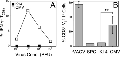
CD8(+) T cells (T(CD8(+))) play a crucial role in immunity to viruses. Antiviral T(CD8(+)) are initially activated by recognition of major histocompatibility complex (MHC) class I-peptide complexes on the surface of professional antigen-presenting cells (pAPC). Migration of pAPC from the site of infection to secondary lymphoid organs is likely required during a natural infection. Migrating pAPC can be directly infected with virus or may internalize antigen derived from virus-infected cells. The use of experimental virus infections to assess the requirement for pAPC migration in initiation of T(CD8(+)) responses has proven difficult to interpret because injected virus can readily drain to secondary lymphoid organs without the need for cell-mediated transport. To overcome this ambiguity, we examined the generation of antigen-specific T(CD8(+)) after immunization with recombinant adenoviruses that express antigen driven by skin-specific or ubiquitous promoters. We show that the induction of T(CD8(+)) in response to tissue-targeted antigen is less efficient than the response to ubiquitously expressed antigen and that the resulting T(CD8(+)) fail to clear all target cells pulsed with the antigenic peptide. This failure to prime a fully functional T(CD8(+)) response results from a reduced period of priming to peripherally expressed antigen versus ubiquitously expressed antigen and correlated with a brief burst of pAPC migration from the skin, a requirement for induction of the response to peripheral antigen. These results indicate that a reduced duration of pAPC migration after virus infection likely reduces the amplitude of the T(CD8(+)) response, allowing persistence of the peripheral virus.
Figures


Similar articles
-
Viral sequestration of antigen subverts cross presentation to CD8(+) T cells.PLoS Pathog. 2009 May;5(5):e1000457. doi: 10.1371/journal.ppat.1000457. Epub 2009 May 29. PLoS Pathog. 2009. PMID: 19478869 Free PMC article.
-
Effective Priming of Herpes Simplex Virus-Specific CD8+ T Cells In Vivo Does Not Require Infected Dendritic Cells.J Virol. 2018 Jan 17;92(3):e01508-17. doi: 10.1128/JVI.01508-17. Print 2018 Feb 1. J Virol. 2018. PMID: 29142130 Free PMC article.
-
Peptide-MHC-I from Endogenous Antigen Outnumber Those from Exogenous Antigen, Irrespective of APC Phenotype or Activation.PLoS Pathog. 2015 Jun 24;11(6):e1004941. doi: 10.1371/journal.ppat.1004941. eCollection 2015 Jun. PLoS Pathog. 2015. PMID: 26107264 Free PMC article.
-
Viral interference with antigen presentation.Nat Immunol. 2002 Nov;3(11):1019-25. doi: 10.1038/ni1102-1019. Nat Immunol. 2002. PMID: 12407410 Review.
-
Regulation of antiviral CD8 T-cell responses.Crit Rev Immunol. 2013;33(6):477-88. doi: 10.1615/critrevimmunol.2013007909. Crit Rev Immunol. 2013. PMID: 24266346 Free PMC article. Review.
Cited by
-
Improving Tumor Retention of Effector Cells in Adoptive Cell Transfer Therapies by Magnetic Targeting.Pharmaceutics. 2020 Aug 27;12(9):812. doi: 10.3390/pharmaceutics12090812. Pharmaceutics. 2020. PMID: 32867162 Free PMC article. Review.
-
Vaccinia Virus: Mechanisms Supporting Immune Evasion and Successful Long-Term Protective Immunity.Viruses. 2024 May 29;16(6):870. doi: 10.3390/v16060870. Viruses. 2024. PMID: 38932162 Free PMC article. Review.
-
Dynamic metabolic reprogramming in dendritic cells: An early response to influenza infection that is essential for effector function.PLoS Pathog. 2020 Oct 26;16(10):e1008957. doi: 10.1371/journal.ppat.1008957. eCollection 2020 Oct. PLoS Pathog. 2020. PMID: 33104753 Free PMC article.
-
Prolonged antigen presentation following an acute virus infection requires direct and then cross-presentation.J Immunol. 2014 Oct 15;193(8):4169-77. doi: 10.4049/jimmunol.1302565. Epub 2014 Sep 15. J Immunol. 2014. PMID: 25225666 Free PMC article.
-
A systemic macrophage response is required to contain a peripheral poxvirus infection.PLoS Pathog. 2017 Jun 14;13(6):e1006435. doi: 10.1371/journal.ppat.1006435. eCollection 2017 Jun. PLoS Pathog. 2017. PMID: 28614386 Free PMC article.
References
-
- Allan, R. S., C. M. Smith, G. T. Belz, A. L. van Lint, L. M. Wakim, W. R. Heath, and F. R. Carbone. 2003. Epidermal viral immunity induced by CD8α+ dendritic cells but not by Langerhans cells. Science 301:1925-1928. - PubMed
-
- Allan, R. S., J. Waithman, S. Bedoui, C. M. Jones, J. A. Villadangos, Y. Zhan, A. M. Lew, K. Shortman, W. R. Heath, and F. R. Carbone. 2006. Migratory dendritic cells transfer antigen to a lymph node-resident dendritic cell population for efficient CTL priming. Immunity 25:153-162. - PubMed
-
- Azukizawa, H., H. Kosaka, S. Sano, W. R. Heath, I. Takahashi, X. H. Gao, Y. Sumikawa, M. Okabe, K. Yoshikawa, and S. Itami. 2003. Induction of T-cell-mediated skin disease specific for antigen transgenically expressed in keratinocytes. Eur. J. Immunol. 33:1879-1888. - PubMed
-
- Bedoui, S., P. G. Whitney, J. Waithman, L. Eidsmo, L. Wakim, I. Caminschi, R. S. Allan, M. Wojtasiak, K. Shortman, F. R. Carbone, A. G. Brooks, and W. R. Heath. 2009. Cross-presentation of viral and self antigens by skin-derived CD103+ dendritic cells. Nat. Immunol. 10:488-495. - PubMed
-
- Brossart, P., A. W. Goldrath, E. A. Butz, S. Martin, and M. J. Bevan. 1997. Virus-mediated delivery of antigenic epitopes into dendritic cells as a means to induce CTL. J. Immunol. 158:3270-3276. - PubMed
Publication types
MeSH terms
Substances
Grants and funding
LinkOut - more resources
Full Text Sources
Research Materials
