. 2010 Mar;16(3):516-28.
doi: 10.1261/rna.2030510. Epub 2010 Jan 20.

Release of SF3 from the intron branchpoint activates the first step of pre-mRNA splicing

Affiliations

Release of SF3 from the intron branchpoint activates the first step of pre-mRNA splicing

Rea M Lardelli et al. RNA. 2010 Mar.

Abstract

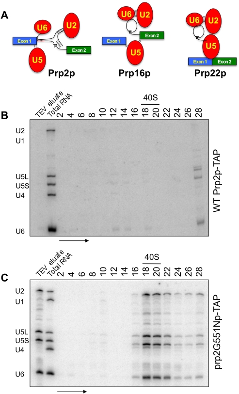
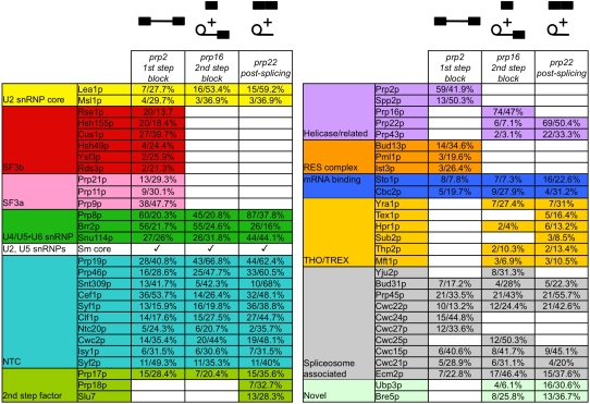
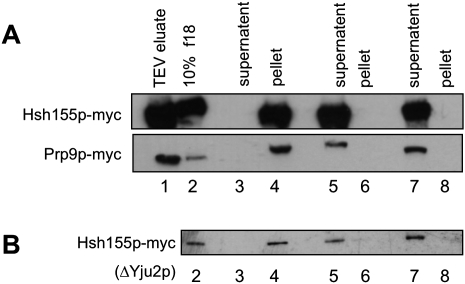
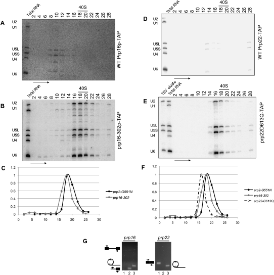
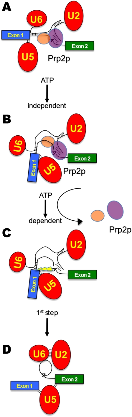
Eukaryotic pre-mRNA splicing is a complex process requiring the precise timing and action of >100 trans-acting factors. It has been known for some time that the two steps of splicing chemistry require three DEAH-box RNA helicase-like proteins; however, their mechanism of action at these steps has remained elusive. Spliceosomes arrested in vivo at the three helicase checkpoints were purified, and first step-arrested spliceosomes were functionally characterized. We show that the first step of splicing requires a novel ATP-independent conformational change. Prp2p then catalyzes an ATP-dependent rearrangement displacing the SF3a and SF3b complexes from the branchpoint within the spliceosome. We propose a model in which SF3 prevents premature nucleophilic attack of the chemically reactive hydroxyl of the branchpoint adenosine prior to the first transesterification. When the spliceosome attains the proper conformation and upon the function of Prp2p, SF3 is displaced from the branchpoint allowing first step chemistry to occur.


Figures

FIGURE 1.
FIGURE 1.
Purification of the first step arrested spliceosome. (A) Graphical representation of the Prp2p- Prp16p- and Prp22p-arrested and -associated spliceosomes purified in this work. RNA from glycerol sedimentation gradients corresponding to affinity purified (B) Prp2p-TAP and (C) prp2G551Np-TAP-associated material were Northern blotted for spliceosomal snRNAs. Splicing complexes are present only in the temperature-shifted mutant corresponding to the U2, U5, and U6 snRNAs sedimenting at ∼40S. Ribosomal subunits were used to determine the peak area of 40S subunit sedimentation. The arrow indicates the direction of sedimentation. Positions of U1, U2, U4, U5, and U6 snRNAs are shown.
FIGURE 2.
FIGURE 2.
Mass spectrometry analysis of the associated proteins from first step-arrested, second step-arrested, and post-second step spliceosomes. Polypeptides corresponding to peak fractions from Figure 1C (prp2-arrested first step), Figure 5B (prp16-arrested second step), from Figure 5E (prp22-arrested spliceosomes) were analyzed by MudPIT. Proteins are categorized by group or functional association. Data for the number of peptides identified and percentage polypeptide sequenced are shown. Polypeptide contaminants are listed in Supplemental Table S1.
FIGURE 3.
FIGURE 3.
Functional characterization of the first step-arrested spliceosome. (A) Metabolic depletion of Yju2p from prp2G551N cells. Western blot analysis of GAL-HA-Yju2p through a time-course in galactose (left) or after shift to dextrose (right). Affinity-purified spliceosomes depleted for Yju2p and arrested at the first step of splicing using the prp2G551N mutation were treated at NPT with no added nucleotide (B), treated at PT with no added nucleotide (C), treated at PT with added ATP (D), or treated at PT with AMP-PNP (E). Proteins in the gradient fractions were subjected to Western blot analysis for Prp9-myc. (F) Prp9p-myc signal was quantitated in gradient fractions from B–E, normalized and graphed (top panel). RNAs from the corresponding fractions were Northern blotted for snRNAs, which were quantitated, normalized, and graphed (bottom panel). (G) RT-PCR analysis of unspliced and partially spliced pre-mRNAs contained in the peak gradient fractions from B, C, and E. Lane 1 of each set is designed to amplify RPP1B pre-mRNA and mRNA. Lane 2 of each set amplifies the RPP1B 3′ splice site junction. Lane 3 of each set specifically amplifies the ACT1 lariat structure using a splint oligonucleotide that bridges the intron sequences of the 5′ splice site and upstream of the branchpoint adenosine and another oligonucleotide that allows for specific amplification within the lariat structure.
FIGURE 4.
FIGURE 4.
SF3a and SF3b are released from the spliceosome concurrent with Prp2p action. (A) Affinity-purified spliceosomes were pelleted through glycerol cushions without splicing (lanes 3,4), with splicing (lanes 5,6), or after disruption with salt (lanes 7,8). Total proteins from supernatant and pellet fractions were Western blotted for the myc tag. (B) Same experiment performed for Hsh155p-myc in A using affinity-purified ΔYju2p spliceosomes. Lane 1 contains 2% of the TEV eluates and lane 2 contains 10% of the 18th fraction from the reactive experiments.
FIGURE 5.
FIGURE 5.
Purification of the prp16- and prp22-arrested spliceosomes. RNA from glycerol sedimentation gradients corresponding to affinity purified (A) Prp16p-TAP and (B) prp16-302p-TAP-associated material were Northern blotted for spliceosomal snRNAs. Splicing complexes are present only in the temperature-shifted mutant corresponding to the U2, U5, and U6 snRNAs sedimenting at ∼40S. (C) Sedimentation differences between first and second step-arrested spliceosomes were calculated using normalized snRNA signals present in the respective gradient fractions. (D) Prp22p-TAP and (E) prp22-G613Qp-TAP-associated material were Northern blotted for snRNAs as in A and B. (F) Graphical representation of sedimentation of prp22-arrested spliceosomes compared to those presented in C. (G) RT-PCR analysis of splicing intermediates and products from affinity purified, gradient isolated prp16- and prp22-arrested spliceosomes. First-step products are seen in prp16-arrested spliceosomes. Spliced mRNA and only the lariat intron are present in prp22-arrested spliceosomes. Lanes are as in Figure 3G.
FIGURE 6.
FIGURE 6.
Model for the activation of the first chemical step of pre-mRNA splicing. (A) The SF3b complex (purple oval) resides on the branchpoint/U2 RNA duplex, while SF3a (orange oval) resides just upstream of SF3b during spliceosome assembly and until the ATP-dependent action of Prp2p. The pre-mRNA is denoted by the green and blue exon boxes and the bottom RNA strand. snRNPs are shown as the red ovals with U2 and U6 snRNAs forming duplexes with each other and the pre-mRNA. U5 snRNP contacts the 5′ splice site. (B) After addition of Prp2p, an ATP-independent rearrangement concomitant with a structural rearrangement occurs. (C) Upon ATP hydrolysis by Prp2p, the SF3a and SF3b components are displaced from the pre-mRNA, uncovering the reactive branchpoint adenosine hydroxyl, which is now properly positioned for in-line attack of the 5′ splice site. (D) Transesterification releases exon1 and the lariat intermediate, formed by the 2′–5′ linkage between the branchpoint and the 5′ splice site.

References

    1. Abovich N, Legrain P, Rosbash M. The yeast PRP6 gene encodes a U4/U6 small nuclear ribonucleoprotein particle (snRNP) protein, and the PRP9 gene encodes a protein required for U2 snRNP binding. Mol Cell Biol. 1990;10:6417–6425. - PMC - PubMed
    1. Abruzzi KC, Lacadie S, Rosbash M. Biochemical analysis of TREX complex recruitment to intronless and intron-containing yeast genes. EMBO J. 2004;23:2620–2631. - PMC - PubMed
    1. Achsel T, Brahms H, Kastner B, Bachi A, Wilm M, Lührmann R. A doughnut-shaped heteromer of human Sm-like proteins binds to the 3′-end of U6 snRNA, thereby facilitating U4/U6 duplex formation in vitro. EMBO J. 1999;18:5789–5802. - PMC - PubMed
    1. Arenas JE, Abelson JN. Prp43: An RNA helicase-like factor involved in spliceosome disassembly. Proc Natl Acad Sci. 1997;94:11798–11802. - PMC - PubMed
    1. Baker-Brachmann C, Davies A, Cost GJ, Caputo E, Li J, Hieter P, Boeke JD. Designer deletion strains derived from Saccharomyces cerevisiae S288C: A useful set of strains and plasmids for PCR-mediated gene disruption and other applications. Yeast. 1998;14:115–132. - PubMed

Publication types

MeSH terms

LinkOut - more resources