. 2010 Apr;298(4):R989-96.
doi: 10.1152/ajpregu.00566.2009. Epub 2010 Jan 20.

The direct molecular effects of fatigue and myosin regulatory light chain phosphorylation on the actomyosin contractile apparatus

Affiliations

The direct molecular effects of fatigue and myosin regulatory light chain phosphorylation on the actomyosin contractile apparatus

Michael J Greenberg et al. Am J Physiol Regul Integr Comp Physiol. 2010 Apr.

Abstract

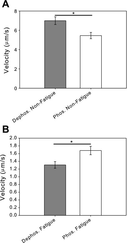
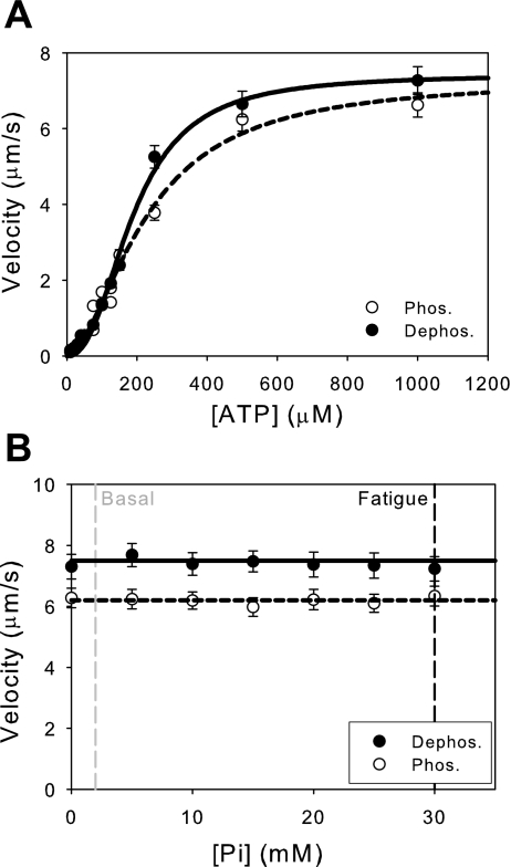
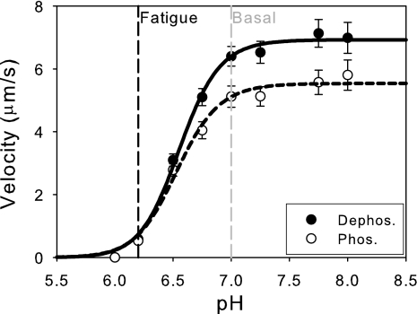
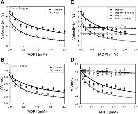
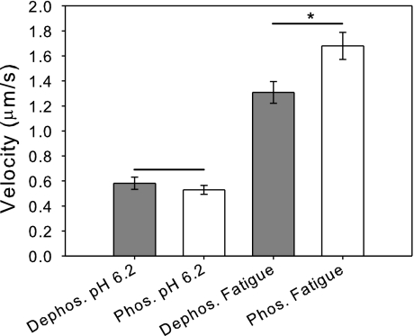
Skeletal muscle, during periods of exertion, experiences several different fatigue-based changes in contractility, including reductions in force, velocity, power output, and energy usage. The fatigue-induced changes in contractility stem from many different factors, including alterations in the levels of metabolites, oxidative damage, and phosphorylation of the myosin regulatory light chain (RLC). Here, we measured the direct molecular effects of fatigue-like conditions on actomyosin's unloaded sliding velocity using the in vitro motility assay. We examined how changes in ATP, ADP, P(i), and pH affect the ability of the myosin to translocate actin and whether the effects of each individual molecular species are additive. We found that the primary causes of the reduction in unloaded sliding velocity are increased [ADP] and lowered pH and that the combined effects of the molecular species are nonadditive. Furthermore, since an increase in RLC phosphorylation is often associated with fatigue, we examined the differential effects of myosin RLC phosphorylation and fatigue on actin filament velocity. We found that phosphorylation of the RLC causes a 22% depression in sliding velocity. On the other hand, RLC phosphorylation ameliorates the slowing of velocity under fatigue-like conditions. We also found that phosphorylation of the myosin RLC increases actomyosin affinity for ADP, suggesting a kinetic role for RLC phosphorylation. Furthermore, we showed that ADP binding to skeletal muscle is load dependent, consistent with the existence of a load-dependent isomerization of the ADP bound state.


Figures

Fig. 1.
Fig. 1.
Fatigue-like conditions reduce actin filament sliding velocity. A: comparison of the unloaded sliding velocities of regulatory light-chain dephosphorylated and phosphorylated myosins under basal conditions. The velocity of phosphorylated myosin (V = 5.5 ± 0.3 μm/s) is significantly reduced compared with the dephosphorylated myosin (V = 7.0 ± 0.4 μm/s ; *P < 0.005). B: comparison of the unloaded sliding velocities of dephosphorylated and phosphorylated myosins under fatigue-like conditions shows that fatigue-like conditions cause a reduction of sliding velocity for both dephosphorylated and phosphorylated myosins. In contrast to the results under basal conditions, the velocity of phosphorylated myosin (V = 1.7 ± 0.1 μm/s) is significantly increased compared with dephosphorylated myosin (V = 1.3 ± 0.1 μm/s; *P < 0.02).
Fig. 2.
Fig. 2.
Fatigue-like reductions in the concentration of ATP and phosphate have little effect on sliding velocity. A: effects of ATP concentration on unloaded sliding velocity. Reducing the available concentration of ATP from basal (5 mM) to fatigue-like conditions (3 mM) has no effect on sliding velocity (data not shown). Also, there is no significant difference in the KM between phosphorylated (KM = 225 ± 25 μM) and dephosphorylated myosins (KM = 190 ± 10 μM; P = 0.21). Data fit to the Hill equation (see text for details). B: effects of changing the concentration of Pi on unloaded sliding velocity. A line with zero slope was fit to the data for illustrative purposes. The addition of Pi has little effect on the unloaded sliding velocity of either phosphorylated or dephosphorylated myosins. Thus, the accumulation of Pi from basal levels (2 mM) to fatigue-like levels (30 mM) has little effect on unloaded sliding velocity.
Fig. 3.
Fig. 3.
Acidic conditions decrease actin filament velocity. Changing the pH from basal levels (7.0) to fatigue-like levels (6.2) causes a significant reduction in sliding velocity. The data were fit to the Hill equation, and it was shown that there is no difference in the pH50 for either phosphorylated (pH50 = 6.54 ± 0.03) or dephosphorylated myosins (pH50 = 6.55 ± 0.02; P = 0.54).
Fig. 4.
Fig. 4.
Exogenous ADP decreases actin filament velocity. The unnormalized (A) and normalized (B) data are shown with fits to a competitive inhibitor model (Eq. 2). Changing the concentration of ADP from basal (0.02) to fatigue-like levels (0.3 mM) causes a significant depression in unloaded sliding velocity for both phosphorylated and dephosphorylated myosins. The inhibition constant for phosphorylated myosin (KI = 95 ± 10 μM) is significantly higher than dephosphorylated myosin (KI = 200 ± 30 μM; P < 0.01), demonstrating that phosphorylated myosin has an approximately 2-fold higher affinity for ADP than dephosphorylated myosin. C and D: load decreases the sensitivity of actin filament velocity to exogenously added ADP. A frictional load was introduced into the motility assay using the low-affinity actin binding protein, α-actinin. The unormalized (C) and normalized (D) data were fit to Eq. 2. Loading the myosin causes a significant reduction in the ability of ADP to depress actomyosin sliding. Loading both the phosphorylated (KI = 6 ± 4 mM) and dephosphorylated (KI = 5 ± 2 mM) myosins causes a lowering of the affinity for exogenously added ADP.
Fig. 5.
Fig. 5.
The combined effects of the individual molecular species involved in fatigue are nonadditive. Fatigue-like changes in pH (6.2) alone causes a significant depression in sliding velocity in the absence of other fatigue-like ionic molecular species. At pH 6.2, there is no significant difference in the unloaded sliding velocity between phosphorylated and dephosphorylated myosins (P = 0.40). On the other hand, the combined effect of all of the molecular species together (at pH 6.2) has an increased velocity compared with acidosis alone. This suggests that effects of the individual molecular species involved in fatigue are nonadditive. Furthermore, phosphorylated myosin has a greater recovery of velocity than dephosphorylated myosin (*P < 0.05) under fatigue-like conditions compared with the effects of acidosis alone.

Similar articles

Cited by

References

    1. Baker JE, Brosseau C, Joel PB, Warshaw DM. The biochemical kinetics underlying actin movement generated by one and many skeletal muscle myosin molecules. Biophys J 82: 2134–2147, 2002 - PMC - PubMed
    1. Bergstrom M, Hultman E. Energy cost and fatigue during intermittent electrical stimulation of human skeletal muscle. J Appl Physiol 65: 1500–1505, 1988 - PubMed
    1. Bing W, Fraser ID, Marston SB. Troponin I and troponin T interact with troponin C to produce different Ca2+-dependent effects on actin-tropomyosin filament motility. Biochem J 327: 335–340, 1997 - PMC - PubMed
    1. Bing W, Knott A, Marston SB. A simple method for measuring the relative force exerted by myosin on actin filaments in the in vitro motility assay: evidence that tropomyosin and troponin increase force in single thin filaments. Biochem J 350: 693–699, 2000 - PMC - PubMed
    1. Butler TM, Siegman MJ, Mooers SU, Barsotti RJ. Myosin light chain phosphorylation does not modulate cross-bridge cycling rate in mouse skeletal muscle. Science 220: 1167–1169, 1983 - PubMed

Publication types