

A comparison of the diagnostic accuracy of in vivo and in vitro photostimulable phosphor digital images in the detection of occlusal caries lesions
- PMID: 20089739
- PMCID: PMC3520404
- DOI: 10.1259/dmfr/91657756
A comparison of the diagnostic accuracy of in vivo and in vitro photostimulable phosphor digital images in the detection of occlusal caries lesions
Abstract
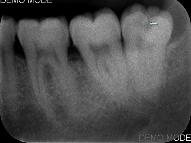
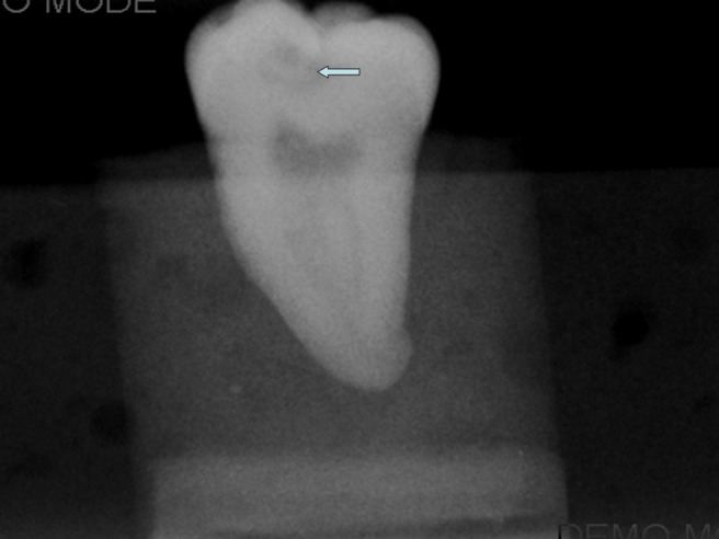
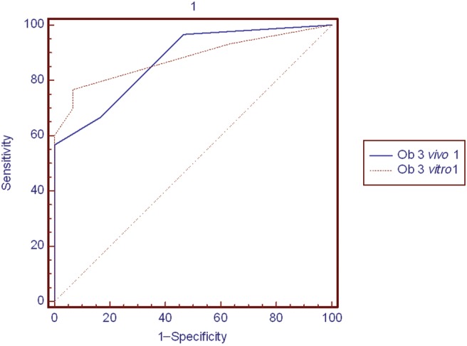
Objectives: The purpose of this study was to compare the accuracy of diagnoses of occlusal caries lesions from digital images captured using a photostimulable phosphor (PSP) sensor under in vivo and in vitro conditions and to present useful clinical data regarding the clinical application of the system.
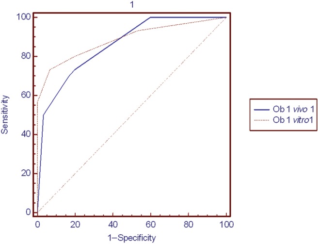
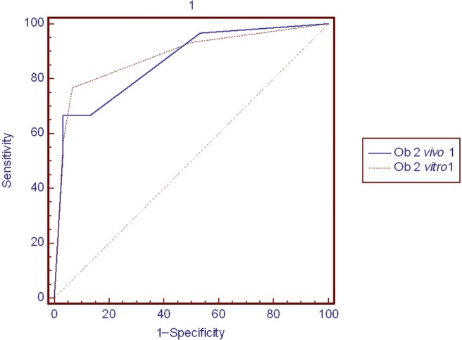
Methods: The study sample comprised 60 mandibular third molars (30 sound and 30 with occlusal caries) requiring extraction. A pre-extraction and post-extraction image of each tooth were acquired using a PSP sensor. A stopwatch was used to record the time required for the clinical procedures. Patient comfort or discomfort during image acquisition was also recorded. Images were evaluated twice by three observers using a five-point scale. Kappa coefficients were calculated to assess intra- and interobserver agreement. Receiver operating characteristic (ROC) curves were used to assess the diagnostic performance of each observer for both in vivo and in vitro images. The t-test was used to compare A(z) values, with a significance level set at 0.05 (alpha _ 0.05). The time required for clinical imaging procedures in patients who claimed discomfort and in those who did not was compared using the Mann-Whitney U-test.
Results: Intraobserver agreement was almost perfect, whereas interobserver agreement was fair to moderate. No statistically significant differences were found in the accuracy of diagnoses of occlusal caries lesions using in vivo and in vitro digital images. The median time needed for image exposure was 1.04 min and the median time needed to complete the image acquisition procedure was 1.45 min.
Conclusions: The diagnosis of accuracy of occlusal caries lesions using in vivo and in vitro digital images yielded similar results.
Figures
Similar articles
-
Intraoral versus extraoral bitewing radiography in detection of enamel proximal caries: an ex vivo study.Dentomaxillofac Radiol. 2016;45(4):20150326. doi: 10.1259/dmfr.20150326. Epub 2016 Feb 19. Dentomaxillofac Radiol. 2016. PMID: 26892946 Free PMC article.
-
A comparative study of different radiographic methods for detecting occlusal caries lesions.Caries Res. 2014;48(6):566-74. doi: 10.1159/000357596. Epub 2014 Jul 29. Caries Res. 2014. PMID: 25073755
-
The effect of delayed scanning of storage phosphor plates on occlusal caries detection.Dentomaxillofac Radiol. 2012 May;41(4):309-15. doi: 10.1259/dmfr/12935491. Epub 2012 Jan 26. Dentomaxillofac Radiol. 2012. PMID: 22282506 Free PMC article.
-
Occlusal caries detection by using a cone-beam CT with different voxel resolutions and a digital intraoral sensor.Oral Surg Oral Med Oral Pathol Oral Radiol Endod. 2010 May;109(5):e63-9. doi: 10.1016/j.tripleo.2009.12.048. Oral Surg Oral Med Oral Pathol Oral Radiol Endod. 2010. PMID: 20416522
-
Digital radiography and caries diagnosis.Dentomaxillofac Radiol. 1998 Jan;27(1):3-11. doi: 10.1038/sj.dmfr.4600321. Dentomaxillofac Radiol. 1998. PMID: 9482015 Review.
Cited by
-
Occlusal caries depth measurements obtained by five different imaging modalities.J Digit Imaging. 2011 Oct;24(5):804-13. doi: 10.1007/s10278-010-9355-9. J Digit Imaging. 2011. PMID: 21116675 Free PMC article.
-
Effect of tube potential and image receptor on the detection of natural proximal caries in primary teeth.Clin Oral Investig. 2011 Dec;15(6):901-7. doi: 10.1007/s00784-010-0461-3. Epub 2010 Sep 14. Clin Oral Investig. 2011. PMID: 20838834
-
Accuracy of conventional and digital radiography in detecting external root resorption.Iran Endod J. 2014 Fall;9(4):241-5. Epub 2014 Oct 7. Iran Endod J. 2014. PMID: 25386202 Free PMC article.
-
Clinical Performance of Diagnostic Methods in Third Molar Teeth with Early Occlusal Caries.Diagnostics (Basel). 2023 Jan 12;13(2):284. doi: 10.3390/diagnostics13020284. Diagnostics (Basel). 2023. PMID: 36673093 Free PMC article.
-
Clinical comparison of intraoral CMOS and PSP detectors in terms of time efficiency, patient comfort, and subjective image quality.Imaging Sci Dent. 2022 Mar;52(1):93-101. doi: 10.5624/isd.20210241. Epub 2022 Feb 11. Imaging Sci Dent. 2022. PMID: 35387105 Free PMC article.
References
-
- Ludlow JB, Mol A. Digital imaging. In: White SC, Pharoah MJ (eds). Oral radiology: Principles and interpretation (5th ed). St. Louis: Mosby, 2004, pp 225–244
-
- Hildebolt CF, Couture RA, Whiting BR. Dental photostimulable phosphor radiography. Dent Clin North Am 2000;44:273–297 - PubMed
-
- Ramamurthy R, Canning CF, Scheetz JP, Farman AG. Time and motion study: a comparison of two photostimulable phosphor imaging systems used in dentistry. Dentomaxillofac Radiol 2006;35:315–318 - PubMed
-
- Farman AG, Farman TT. A comparison of 18 different x-ray detectors currently used in dentistry. Oral Surg Oral Med Oral Pathol Oral Radiol Endod 2005;99:485–489 - PubMed
-
- Hintze H, Wenzel A, Frydenberg M. Accuracy of caries detection with four storage phosphor systems and E speed radiographs. Dentomaxillofac Radiol 2002;31:170–175 - PubMed
Publication types
MeSH terms
LinkOut - more resources
Full Text Sources
Medical
Miscellaneous
