

Modulation transfer function evaluation of cone beam computed tomography for dental use with the oversampling method
- PMID: 20089741
- PMCID: PMC3520411
- DOI: 10.1259/dmfr/27069629
Modulation transfer function evaluation of cone beam computed tomography for dental use with the oversampling method
Abstract
Objectives: The aim was to investigate the possibility of evaluating the modulation transfer function (MTF) of cone beam CT (CBCT) for dental use using the oversampling method.
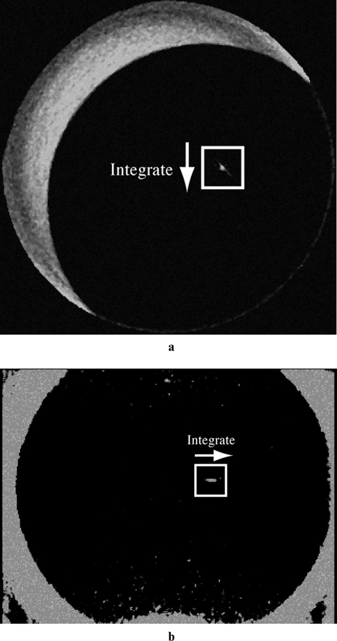
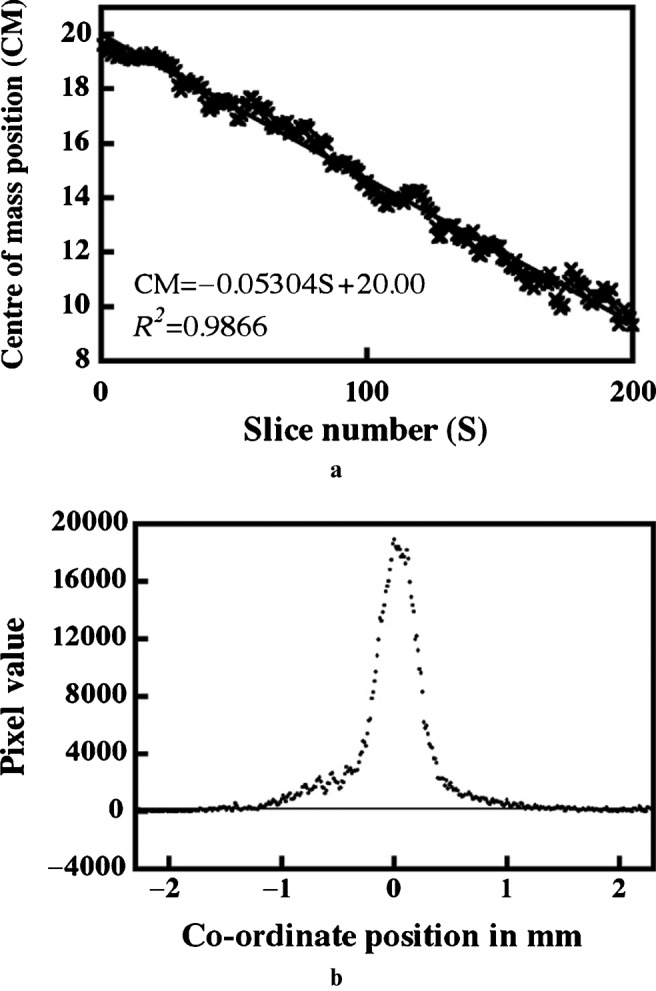
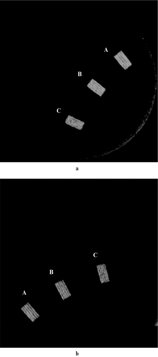
Methods: The CBCT apparatus (3D Accuitomo) with an image intensifier was used with a 100 mum tungsten wire placed inside the scanner at a slight angle to the plane perpendicular to the plane of interest and scanned. 200 contiguous reconstructed images were used to obtain the oversampling line-spread function (LSF). The MTF curve was obtained by computing the Fourier transformation from the oversampled LSF. Line pair tests were also performed using Catphan(R).
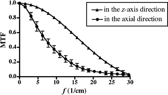
Results: The oversampling method provided smooth and reproducible MTF curves. The MTF curves revealed that the spatial resolution in the z-axis direction was significantly higher than that in the axial direction. This result was also confirmed by the line pair test.
Conclusions: MTF analysis was performed successfully using the oversampling method. In addition, this study clarified that the 3D Accuitomo had high spatial resolution, especially in the z-axis direction.
Figures
References
-
- Tantanapornkul W, Okouchi K, Fujiwara Y, Yamashiro M, Maruoka Y, Ohbayashi N., et al A comparative study of cone-beam computed tomography and conventional panoramic radiography in assessing the topographic relationship between the mandibular canal and impacted third molars. Dentomaxillofac Radiol 2007;103:253–259 - PubMed
-
- Momin MA, Okouchi K, Watanabe H, Imaizumi A, Omura K, Amagasa T, et al. Diagnostic accuracy of cone-beam CT in the assessment of mandibular invasion of lower gingival carcinoma: comparison with conventional panoramic radiography. Eur J Radiol 2009;72:75–81 - PubMed
-
- Loubele M, Guerrero ME, Jacobs R, Suetens P, van Steenberghe D. A comparison of jaw dimensional and quality assessments of bone characteristics with cone-beam CT, spiral tomography, and multi-slice spiral CT. Int J Oral Maxillofac Implants 2007;22:446–454 - PubMed
-
- Arai Y, Tammisalo E, Hashimoto K, Shinoda K. Development of a compact computed tomographic apparatus for dental use. Dentomaxillofac Radiol 1999;28:245–248 - PubMed
-
- Araki K, Maki K, Seki K, Sakamaki K, Harata Y, Sakaino R, et al. Characteristics of a newly developed dentomaxillofacial X-ray cone beam CT scanner (CB MercuRay): system configuration and physical properties. Dentomaxillofac Radiol 2004;33:51–59 - PubMed
Publication types
MeSH terms
LinkOut - more resources
Full Text Sources
Miscellaneous
