. 2010 Mar;17(3):342-53.
doi: 10.1128/CVI.00347-09. Epub 2010 Jan 20.

Nasolacrimal duct closure modulates ocular mucosal and systemic CD4(+) T-cell responses induced following topical ocular or intranasal immunization

Affiliations

Nasolacrimal duct closure modulates ocular mucosal and systemic CD4(+) T-cell responses induced following topical ocular or intranasal immunization

Aziz Alami Chentoufi et al. Clin Vaccine Immunol. 2010 Mar.

Abstract

Both topical ocular and topical intranasal immunizations have been reported to stimulate the ocular mucosal immune system (OMIS) and the systemic immune system. Nasolacrimal ducts (NLDs) are the connecting bridges between the OMIS and nasal cavity-associated lymphoid tissue (NALT). These ducts drain topical ocularly administrated solutions into the inferior meatus of the nose to reach the NALT. Inversely, NLDs also drain intranasally administrated solutions to the mucosal surface of the eye and thus the OMIS. This unique anatomical connection between the OMIS and NALT systems provoked us to test whether the OMIS and NALT are immunologically interdependent. In this report, we show that both topical ocular administration and topical intranasal administration of a mixture of immunodominant CD4(+) T-cell epitope peptides from herpes simplex virus type 1 (HSV-1) glycoprotein D (gD) emulsified with the CpG(2007) mucosal adjuvant are capable of inducing local (in conjunctiva) as well as systemic (in spleen) HSV-peptide-specific CD4(+) T-cell responses. Interestingly, surgical closure of NLDs did not significantly alter local ocular mucosal CD4(+) T-cell responses induced following topical ocular immunization but did significantly enhance systemic CD4(+) T-cell responses (as measured by both T-cell proliferation and gamma interferon (IFN-gamma) production; P < 0.005). In contrast, NLD closure significantly decreased ocular mucosal, but not systemic, CD4(+) T-cell responses following intranasal administration of the same vaccine solution (P < 0.001). The study suggests that NALT and the OMIS are immunologically interconnected.


Figures

FIG. 1.
FIG. 1.
Schematic representation of rabbit nasolacrimal system. the nasolacrimal canal is shown open (A) or closed (B) as described in Methods. The two sharp bends, the proximal maxillary bend (pb) and the bend at incisor tooth (ib), are indicated. The nasolacrimal canal system is composed of the lacrimal canaliculi (Lc), lacrimal sac (Ls), nasolacrimal duct (Ld), and the nasal meatus (n).
FIG. 2.
FIG. 2.
Schematic representation of CD4+ T-cell epitope peptides identified from HSV-1 gD. The amino acid sequence in a single-letter code and position of 12 HSV-1 (strain 17) gD epitope peptides is illustrated. Black box: the transmembrane domain of HSV-1 gD and no peptides was identified in this region. Ten peptides belong to the external N-terminal portion (gray box) of gD (gD1-29, gD22-52, gD49-82, gD77-104, gD96-123, gD121-152, gD146-179, gD176-206, gD200-234, and gD228-257), one lies adjacent to the hydrophobic membrane anchor domain of gD (gD287-317), and one is part of the proposed hydrophilic C-terminal cytoplasmic portion of gD (gD332-358) (white box). It should be emphasized that of the 12 predicted regions, six mapped to nonglycosylated sites of gD (gD1-29, gD49-82, gD146-179, gD228-257, gD287-317, and gD332-358). The sequences of the four-immunodominant peptide CD4+ T-cell epitopes are highlighted in bold.
FIG. 3.
FIG. 3.
Topical ocular immunization with gD peptides plus CpG2700 induces local and systemic Ag-specific T cells and protects against HSV-1 replication in the eyes of rabbits. Four groups of rabbits (10 per group) were immunized three times topically on the eyes (black bar) with the mixture of four peptides plus CpG2007 adjuvant, as described in Materials and Methods. Group G1 received gD1-29, gD22-52, gD77-104, and gD121-152; Group G2 received gD49-82, gD146-179, gD200-234, and gD287-317; Group G3 received gD96-123, gD176-206, gD228-257, and gD332-358; and Group G4 received CpG2007 alone (white bar, nonimmunized control). Fourteen days after the third immunization, conjunctiva (A) or spleen (B) was harvested from each animal and local (in conjunctiva) and systemic (in spleen) T cells were restimulated for 96 h in vitro with a mixture of the immunizing gD peptides or left unstimulated (None). T-cell proliferations were determined by [3H]thymidine incorporation after an additional 16 h of stimulation. The P values compare immunized versus nonimmunized animals using an ANOVA test. *, P < 0.05. (C) Another four groups of rabbits (10 per group) were immunized three times topically on the eyes (black bar) with the mixture of four peptides plus CpG2007 adjuvant, as above. Ten days after the third immunization, all rabbits were ocularly challenged with 2 × 10⁵ PFU/eye of HSV-1 (McKrae). To collect the tears, both eyes were swabbed once daily (from day 1 to day 10 postinfection) with a Dacron swab and transferred immediately to a 75-mm culture tube containing 0.5 ml of medium. The presence of HSV-1 in both eyes was monitored by plaque-forming assay. The data show the average virus titers on the peak day (day 5) of postinfection in rabbits immunized with designated groups of peptides. The results represent the average of results from 2 independent experiments.
FIG. 4.
FIG. 4.
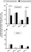
HSV-1 gD peptides induced local and systemic T-cell responses in both topical ocular and nasal immunization setups. Rabbits (n = 5) were immunized either ocularly (gray bars) or nasally (black bars) with a mixture of 4 immunodominant CD4+ T-cell epitopes from HSV-1 glycoprotein gD (25 μg of each gD peptide, gD49-82, gD146-179, gD228-257, and gD332-358) and 25 μg of CpG (adjuvant) three times every 14 days in a total volume of 50 μl and delivered as a drop. Ten days after the last immunization, individual conjunctiva and spleen were harvested and lymphoid cells were isolated. The cells were stained with CFSE (2.5 μM) and stimulated (3 × 10⁵ cell/well) in vitro in the presence or absence of 10 μg/ml of the individual HSV-gD peptides gD49-82, gD146-179, gD228-257 and gD332-358, respectively. The CFSE-labeled cells were harvested, washed, and stained with PE-anti-rabbit CD4 antibody. Dividing cells in conjunctiva (A) or spleen (B) were analyzed by flow cytometry, and the following formula was used to calculate the absolute number of dividing cells in the present study. No. of dividing cells = no. of gated CD4+ CFSElow cells × no. of total CD4+ lymphocytes (i.e., CD4+ CFSElow + CD4+ CFSEhigh lymphocytes)/no. of total gated lymphocytes.
FIG. 5.
FIG. 5.
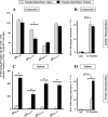
Nasolacrimal duct blockage enhanced the systemic immune response induced by topical ocular immunization with HSV-1 gD peptides. Rabbits (n = 5) with closed (black bars) or open (gray bars) nasolacrimal canals were ocularly immunized with 4 immunodominant CD4+ T-cell epitopes from HSV-1 glycoprotein gD (25 μg of each gD peptide, gD49-82, gD146-179, gD228-257, and gD332-358) and 25 μg of CpG mucosal adjuvant) three times every 14 days as a 50-μl drop per eye. Ten days after the last immunization, individual conjunctiva and spleens were harvested and lymphoid cells were isolated. The cells were stained with CFSE (2.5 μM) and stimulated in vitro (3 × 10⁵ cell/well) in the presence or absence of 10 μg/ml of the individual HSV-gD peptides gD49-82, gD146-179, gD228-257, and gD332-358 for 5 days at 37°C and 5% CO2. The CFSE-labeled cells were harvested, washed, and stained with PE-anti-rabbit CD4 antibody. Cycling cells were analyzed by flow cytometry, and the following formula was used to calculate the absolute number in the present study: no. of dividing cells = no. of gated CD4+ CFSElow cells × no. of total CD4+ lymphocytes (i.e., CD4+ CFSElow + CD4+ CFSEhigh lymphocytes)/no. of total gated lymphocytes. Panels A and C represent the absolute numbers of gD-peptide-specific CD4+ T cells in conjunctiva and spleen, respectively. The amount of IFN-γ transcript produced in the conjunctiva (B) or spleen (D) following topical ocular immunization with gD peptides plus CpG (G2 peptides) or PBS (Mock) was measured by real-time PCR and is represented as an average of results from 2 independent experiments.
FIG. 6.
FIG. 6.
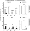
Nasolacrimal-duct blockage abrogates the ocular immune response induced following intranasal immunization with HSV-1 gD peptides. Rabbits (n = 5) with a closed (black bars) or open (gray bars) nasolacrimal canal were intranasally immunized with 4 immunodominant CD4+ T-cell epitopes from HSV-1 glycoprotein gD (25 μg of each of the gD peptides gD49-82, gD146-179, gD228-257, and gD332-358) and 25 μg of CpG mucosal adjuvant three times every 14 days as a 50-μl drop in each nose passage. Ten days after the last immunization, individual conjunctiva and spleen were harvested and lymphoid cells were isolated. The cells were labeled with CFSE (2.5 μM) and stimulated in vitro (3 × 10⁵ cell/well) in the presence or absence of 10 μg/ml of the individual HSV-gD peptides gD49-82, gD146-179, gD287-317, and gD332-358 as designated. The cells were incubated for 5 days at 37°C and 5% CO2. The CFSE-labeled cells were harvested, washed, and stained with PE-anti-rabbit CD4 antibody. The following formula was used to calculate the absolute number of dividing cells: no. of dividing cells = no. of gated CD4+ CFSElow cells × no. of total CD4+ lymphocytes (i.e., CD4+ CFSElow + CD4+ CFSEhigh lymphocytes)/no. of total gated lymphocytes. Panels A and C represent the absolute numbers of proliferated gD-peptide-specific CD4+ T cells in the conjunctiva and spleen, respectively. The amount of IFN-γ transcript produced in the conjunctiva (B) or spleen (D) following topical intranasal immunization with gD peptides plus CpG (G2 peptides) or PBS (Mock) was measured by RT-PCR and represented as an average of results from 2 independent experiments.
FIG. 7.
FIG. 7.
Topical ocular immunization with HSV-1 gD peptides induced infiltration of CD11b+ TLR9+ cells in conjunctiva and spleen. (A) An expression profile of Toll-like receptors in isolated CD11b+ cells from rabbit conjunctiva (0.24 × 10⁶ per assay) were first surface stained with 1 μl of anti-rabbit CD11b-FITC antibody, followed by intracellular staining with 3 μl of either anti-human TLR2-PE, TLR3-PE, TLR4-PE, TLR8-PE, or TLR9-PE in each set. The dotted line in panel A represents the CD11b+ cells stained with isotype control MAbs, and the solid line represents CD11b+ cells stained with anti-human TLR-PE antibody. (B) Level of each TLR in CD11b+ cells as a function of mean fluorescence intensity (MFI). *, P < 0.05 in a comparison of the MFI of TLR2 and TLR9 expression to that for the isotype controls in the CD11b+ populations. (C) Detection of TLR9 in rabbit conjunctiva by Western blotting. In both panels, lane 1 contains rainbow molecular weight markers; lane 2 contains 5 μg of human PBMC lysate; lanes 3 and 4 contain 20 μg of CD11c+ and CD11b+ cell lysate isolated from rabbit conjunctiva. The left membrane was probed with mouse anti-human TLR9 (IgG1-HRP at a 1:500 dilution), and the right membrane was probed with isotype control antibody (anti-mouse IgG1-HRP at a 1:500 dilution).
FIG. 8.
FIG. 8.
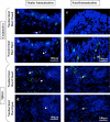
CD11b+ cells expressing TLR9 are mobilized following topical ocular immunization. Detection of CD11b+ TLR9+ cells in the conjunctiva and spleen by immunofluorescence staining is shown. Conjunctiva and spleen tissues from rabbits immunized ocularly or intranasally with gD peptides, with or without NLD closure, were snap-frozen in liquid nitrogen and processed for immunostaining as described in Materials and Methods. The sections were doubly stained with FITC-labeled anti-rabbit CD11b and PE-labeled anti-human TLR9. The sections were counterstained with DAPI for nuclear localization (blue). The arrows show the doubly positive CD11b+ TLR9+ cells as orange to yellow dots.

References

    1. BenMohamed, L., Y. Belkaid, E. Loing, K. Brahimi, H. Gras-Masse, and P. Druilhe. 2002. Systemic immune responses induced by mucosal administration of lipopeptides without adjuvant. Eur. J. Immunol. 32:2274-2281. - PubMed
    1. BenMohamed, L., G. Bertrand, C. D. McNamara, H. Gras-Masse, J. Hammer, S. L. Wechsler, and A. B. Nesburn. 2003. Identification of novel immunodominant CD4+ Th1-type T-cell peptide epitopes from herpes simplex virus glycoprotein D that confer protective immunity. J. Virol. 77:9463-9473. - PMC - PubMed
    1. BenMohamed, L., R. Krishnan, C. Auge, J. F. Primus, and D. J. Diamond. 2002. Intranasal administration of a synthetic lipopeptide without adjuvant induces systemic immune responses. Immunology 106:113-121. - PMC - PubMed
    1. BenMohamed, L., S. L. Wechsler, and A. B. Nesburn. 2002. Lipopeptide vaccines—-yesterday, today, and tomorrow. Lancet Infect. Dis. 2:425-431. - PubMed
    1. Bettahi, I., A. B. Nesburn, S. Yoon, X. Zhang, A. Mohebbi, V. Sue, A. Vanderberg, S. L. Wechsler, and L. BenMohamed. 2007. Protective immunity against ocular herpes infection and disease induced by highly immunogenic self-adjuvanting glycoprotein D lipopeptide vaccines. Invest. Ophthalmol. Vis. Sci. 48:4643-4653. - PubMed

Publication types

MeSH terms