

Reliability and frequency response of excitatory signals transmitted to different types of retinal ganglion cell
- PMID: 20089819
- PMCID: PMC2887626
- DOI: 10.1152/jn.00871.2009
Reliability and frequency response of excitatory signals transmitted to different types of retinal ganglion cell
Abstract
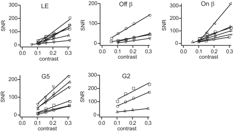
The same visual stimulus evokes a different pattern of neural signals each time the stimulus is presented. Because this unreliability reduces visual performance, it is important to understand how it arises from neural circuitry. We asked whether different types of ganglion cell receive excitatory signals with different reliability and frequency content and, if so, how retinal circuitry contributes to these differences. If transmitter release is governed by Poisson statistics, the SNR of the postsynaptic currents (ratio of signal power to noise power) should grow linearly with quantal rate (qr), a prediction that we confirmed experimentally. Yet ganglion cells of the same type receive quanta at different rates. Thus to obtain a measure of reliability independent of quantal rate, we calculated the ratio SNR/qr, and found this measure to be type-specific. We also found type-specific differences in the frequency content of postsynaptic currents, although types whose dendrites branched at nearby levels of the inner plexiform layer (IPL) had similar frequency content. As a result, there was an orderly distribution of frequency response through the depth of the IPL, with alternating layers of broadband and high-pass signals. Different types of bipolar cell end at different depths of the IPL and provide excitatory synapses to ganglion cell dendrites there. Thus these findings indicate that a bipolar cell synapse conveys signals whose temporal message and reliability (SNR/qr) are determined by neuronal type. The final SNR of postsynaptic currents is set by the dendritic membrane area of a ganglion cell, which sets the numbers of bipolar cell synapses and thus the rate at which it receives quanta [SNR = qr x (SNR/qr)].
Figures


 ), peak (✚), and exponential decay (solid lines). B: sEPSCs were averaged and then integrated to give quantal charge q′. C: average quantal charge for 6 types of retinal ganglion cell.


Similar articles
-
A Mammalian Retinal Ganglion Cell Implements a Neuronal Computation That Maximizes the SNR of Its Postsynaptic Currents.J Neurosci. 2017 Feb 8;37(6):1468-1478. doi: 10.1523/JNEUROSCI.2814-16.2016. Epub 2016 Dec 30. J Neurosci. 2017. PMID: 28039376 Free PMC article.
-
Parallel cone bipolar pathways to a ganglion cell use different rates and amplitudes of quantal excitation.J Neurosci. 2000 Jun 1;20(11):3956-63. doi: 10.1523/JNEUROSCI.20-11-03956.2000. J Neurosci. 2000. PMID: 10818130 Free PMC article.
-
Synaptic noise is an information bottleneck in the inner retina during dynamic visual stimulation.J Physiol. 2014 Feb 15;592(4):635-51. doi: 10.1113/jphysiol.2013.265744. Epub 2013 Dec 2. J Physiol. 2014. PMID: 24297850 Free PMC article.
-
How voltage-gated ion channels alter the functional properties of ganglion and amacrine cell dendrites.Arch Ital Biol. 2002 Oct;140(4):347-59. Arch Ital Biol. 2002. PMID: 12228988 Review.
-
Synaptic activity, visual experience and the maturation of retinal synaptic circuitry.J Physiol. 2008 Sep 15;586(18):4347-55. doi: 10.1113/jphysiol.2008.159202. Epub 2008 Jul 31. J Physiol. 2008. PMID: 18669531 Free PMC article. Review.
Cited by
-
Selective synaptic connections in the retinal pathway for night vision.J Comp Neurol. 2019 Jan 1;527(1):117-132. doi: 10.1002/cne.24313. Epub 2017 Sep 15. J Comp Neurol. 2019. PMID: 28856684 Free PMC article.
-
Cross inhibition from ON to OFF pathway improves the efficiency of contrast encoding in the mammalian retina.J Neurophysiol. 2012 Nov;108(10):2679-88. doi: 10.1152/jn.00589.2012. Epub 2012 Aug 29. J Neurophysiol. 2012. PMID: 22933723 Free PMC article.
-
Convergence and Divergence of CRH Amacrine Cells in Mouse Retinal Circuitry.J Neurosci. 2018 Apr 11;38(15):3753-3766. doi: 10.1523/JNEUROSCI.2518-17.2018. Epub 2018 Mar 23. J Neurosci. 2018. PMID: 29572434 Free PMC article.
-
A Mammalian Retinal Ganglion Cell Implements a Neuronal Computation That Maximizes the SNR of Its Postsynaptic Currents.J Neurosci. 2017 Feb 8;37(6):1468-1478. doi: 10.1523/JNEUROSCI.2814-16.2016. Epub 2016 Dec 30. J Neurosci. 2017. PMID: 28039376 Free PMC article.
-
Why do axons differ in caliber?J Neurosci. 2012 Jan 11;32(2):626-38. doi: 10.1523/JNEUROSCI.4254-11.2012. J Neurosci. 2012. PMID: 22238098 Free PMC article.
References
-
- Amthor FR, Oyster CW, Takahashi ES. Morphology of on-off direction-selective ganglion cells in the rabbit retina. Brain Res 298: 187–190, 1984 - PubMed
-
- Amthor FR, Takahashi ES, Oyster CW. Morphologies of rabbit retinal ganglion cells with complex receptive fields. J Comp Neurol 280: 97–121, 1989 - PubMed
-
- Berson DM, Pu M, Famiglietti EV. The zeta cell: a new ganglion cell type in cat retina. J Comp Neurol 399: 269–288, 1998 - PubMed
Publication types
MeSH terms
Grants and funding
LinkOut - more resources
Full Text Sources
Research Materials