. 2010 Mar 15;21(6):1033-46.
doi: 10.1091/mbc.e09-10-0914. Epub 2010 Jan 20.

Vesicular calcium regulates coat retention, fusogenicity, and size of pre-Golgi intermediates

Affiliations

Vesicular calcium regulates coat retention, fusogenicity, and size of pre-Golgi intermediates

Marvin Bentley et al. Mol Biol Cell. .

Abstract

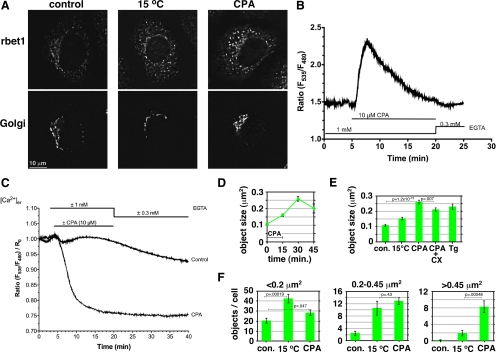
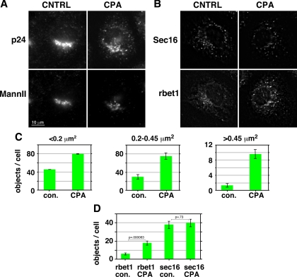
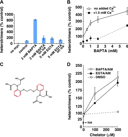
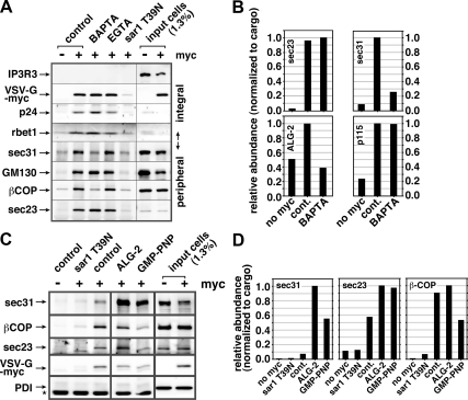
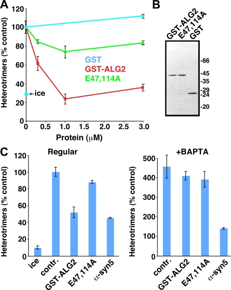
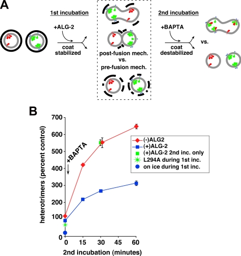
The significance and extent of Ca(2+) regulation of the biosynthetic secretory pathway have been difficult to establish, and our knowledge of regulatory relationships integrating Ca(2+) with vesicle coats and function is rudimentary. Here, we investigated potential roles and mechanisms of luminal Ca(2+) in the early secretory pathway. Specific depletion of luminal Ca(2+) in living normal rat kidney cells using cyclopiazonic acid (CPA) resulted in the extreme expansion of vesicular tubular cluster (VTC) elements. Consistent with this, a suppressive role for vesicle-associated Ca(2+) in COPII vesicle homotypic fusion was demonstrated in vitro using Ca(2+) chelators. The EF-hand-containing protein apoptosis-linked gene 2 (ALG-2), previously implicated in the stabilization of sec31 at endoplasmic reticulum exit sites, inhibited COPII vesicle fusion in a Ca(2+)-requiring manner, suggesting that ALG-2 may be a sensor for the effects of vesicular Ca(2+) on homotypic fusion. Immunoisolation established that Ca(2+) chelation inhibits and ALG-2 specifically favors residual retention of the COPII outer shell protein sec31 on pre-Golgi fusion intermediates. We conclude that vesicle-associated Ca(2+), acting through ALG-2, favors the retention of residual coat molecules that seem to suppress membrane fusion. We propose that in cells, these Ca(2+)-dependent mechanisms temporally regulate COPII vesicle interactions, VTC biogenesis, cargo sorting, and VTC maturation.


Figures

Figure 1.
Figure 1.
Luminal Ca2+ regulates size of rbet1-positive pre-Golgi structures. (A) NRK cells were either mock treated, incubated at 15°C for 30 min, or treated with CPA to deplete luminal Ca2+ (see Materials and Methods), and then fixed and immunostained for rbet1 and the Golgi marker GM130. Shown are single focal planes from deconvolved widefield image stacks. (B) Cytosolic Ca2+ dynamics during the CPA regimen to deplete luminal Ca2+. Fluorescence ratios are calibrated to free Ca2+ values in Supplemental Figure S1A. (C) Luminal ER Ca2+ dynamics during the CPA regimen. Fluorescence ratios are calibrated to free Ca2+ values in Supplemental Figure S1B. (D–F) Quantitation of peripheral rbet1-positive objects in experiments such as described in A. Values represent per cell means derived from at least 20 randomly chosen cells. Error bars display SE. CX, cycloheximide; Tg, thapsigargin. In F, only objects that fall within the size bins indicated above each plot are included in each panel. Selected p values from two-tailed Student's t test are included. Areas of objects were calculated assuming that one image pixel width calibrates to 224 nm in the cell. Single-pixel objects (with calculated area 0.05 μm2) are subresolution but were not eliminated from the size analysis; they did not contribute to the 0.2–0.45 μm2 and >0.45 μm2 size bins in F.
Figure 2.
Figure 2.
Luminal Ca2+ regulates the size of ERGIC but not ERES structures. (A) NRK cells were either mock treated or treated with CPA to deplete luminal Ca2+ (see Materials and Methods), then fixed and immunostained for p24 and the Golgi marker mannosidase II. Shown are single focal planes from deconvolved widefield image stacks. (B) Immunolabeling for rbet1 and sec16 under the same experimental conditions as described in A. (C) Quantitation of numbers of peripheral p24-positive objects in three size bins under the experimental conditions shown in A. (D) Quantitation of peripheral objects positive for rbet1 and sec16 from the experiment shown in B. All size bins were included in one analysis. The rbet1 and sec16 object data are from precisely the same set of double-labeled cells. Values represent means derived from at least 20 randomly chosen cells. Error bars display SE. Selected p values from two-tailed Student's t test are included.
Figure 3.
Figure 3.
BAPTA, but not EGTA, stimulates COPII vesicle fusion. (A) Homotypic COPII vesicle fusion measured using the in vitro VSV-G heterotrimer cargo mixing assay. The no myc control includes radioactive VSV-G* vesicles, but not VSV-G-myc vesicles, to control for the specificity of the anti-myc immunoprecipitation. BAPTA and EGTA were included in fusion reactions at the indicated concentrations. The on ice reactions had the indicated chelators added after the fusion incubation, before detergent solubilization, to control for possible nonfusion-related effects of the chelators. (B) Titration of fusion reactions with BAPTA in the presence and absence of excess free Ca2+. Because the BAPTA stimulation curve is pushed to the right, it seems that only free BAPTA, and not the Ca2+-bound chelator, has a stimulatory effect on heterotrimer formation. (C) Structures of EGTA (black) and BAPTA (black plus red). (D) Effects of aminomethoxy (/AM) derivatives of BAPTA and EGTA on homotypic COPII vesicle fusion. Both /AM esters were able to partially mimic the effects of BAPTA, suggesting that the selective effects of BAPTA were due to its ability to rapidly chelate escaping luminal Ca2+. Fusion assay data are presented as means of duplicate determinations with error bars representing SE where larger than symbol size.
Figure 4.
Figure 4.
Luminal Ca2+ and ALG-2 regulate the retention of select coat subunits on pre-Golgi fusion intermediates. (A) BAPTA specifically extracts select COPI and COPII subunits from forming pre-Golgi intermediates. Homotypic fusion in vitro assays were conducted in the absence or presence of 2 mM BAPTA or EGTA (indicated above), and immunoisolated using anti-myc antibodies. Vesicles were generated from VSV-G-myc transfected cells (+) or nontransfected cells (−) to demonstrate specificity of isolation. In addition, 1 μM purified sar1 T39N was included during the budding stage to demonstrate that the isolated intermediates are COPII derived. Immunoblotted proteins are indicated along left edge. IP3R3, a resident ER membrane protein, additionally demonstrates specificity of budding. (B) Quantitation of a similar immunoisolation experiment (see blot in Supplemental Figure S3). In this experiment, it can be seen that peripheral membrane protein ALG-2 is also sensitive to BAPTA, but p115 is not. (C) Similar immunoisolation experiment where purified ALG-2 is present during the fusion experiment. ALG-2 caused a dramatic retention of outer shell component sec31 but not the COPI component β-COP. The absence of ER resident luminal protein PDI demonstrates specificity of the intermediates isolated. A star indicates the position of cross-reactive antibody heavy chain bands. GMP-PNP shows an estimate of components present on a fully coated vesicle as opposed to components remaining on tethered/fused pre-Golgi intermediates (D) Quantitation of experiment from C. For quantitations (B and D), band intensities were normalized to the recovery of cargo VSV-G-myc in each condition before plotting them on a relative scale with the highest recovery set to 1. Differences in band intensities seen in the input cells lanes (A) were caused by using slightly denser cell suspensions for the nontransfected cell controls.
Figure 5.
Figure 5.
ALG-2 regulates in vitro COPII vesicle homotypic fusion in a Ca2+-dependent manner. (A) Homotypic COPII vesicle fusion measured using the in vitro VSV-G heterotrimer cargo mixing assay. Purified GST, GST-ALG-2 wild-type, or GST-ALG-2 E47,114A mutant proteins were included in fusion incubations at the indicated concentrations. The ice control includes radioactive VSV-G* vesicles and VSV-G-myc vesicles, but the low temperature prevents a specific fusion signal. (B) PAGE gel on purified proteins used in A and C, stained with Coomassie Blue. Proteins were used in fusion assays in the same proportions as on the gel. (C) Fusion experiment using partially inhibitory doses of the proteins indicated along the bottom. The left panel experiment was conducted in the absence of BAPTA. The right panel experiment was conducted, in the same experiment, in the presence of 2 mM BAPTA. Fusion values for both panels are normalized to the positive control signal in the absence of BAPTA (set to 100%). BAPTA negates the specific inhibitory effect of GST-ALG-2 but not that of anti-syntaxin 5 antibody. Fusion assay data are presented as means of duplicate determinations, with error bars representing SE where they are larger than symbol size.
Figure 6.
Figure 6.
ALG-2–dependent coat retention primarily inhibits via a prefusion mechanism. (A) Outline of experiment to distinguish between pre- or postfusion mechanisms by which ALG-2 inhibits heterotrimer assay (see text for explanation). Cargos VSV-G-myc and radioactive VSV-G* are depicted in green and red, respectively. COPII coat is represented by thick black lines. (B) Heterotrimer data from experiment depicted in A. Heterotrimers are normalized with 100% representing the amount produced during the first incubation in the absence of recombinant ALG-2, before addition of BAPTA and second incubation. BAPTA was omitted from the conditions plotted at time 0; BAPTA was added to all other conditions, while on ice, just before beginning the second incubation at 32°C. ALG-2 added during the first incubation had a dramatic consequence on the subsequent heterotrimer signal (red vs. blue lines), but ALG-2 added only during the second incubation had no influence (green square). Plotted are means of duplicate reactions, with error bars to represent SE where they exceeded symbol size.

Similar articles

Cited by

References

    1. Ahluwalia J. P., Topp J. D., Weirather K., Zimmerman M., Stamnes M. A role for calcium in stabilizing transport vesicle coats. J. Biol. Chem. 2001;276:34148–34155. - PubMed
    1. Allan B. B., Moyer B. D., Balch W. E. Rab1 recruitment of p115 into a cis-SNARE complex: programming budding COPII vesicles for fusion. Science. 2000;289:444–448. - PubMed
    1. Alvarez C., Garcia-Mata R., Hauri H. P., Sztul E. The p115-interactive proteins GM130 and giantin participate in endoplasmic reticulum-Golgi traffic. J. Biol. Chem. 2001;276:2693–2700. - PubMed
    1. Amodio G., Renna M., Paladino S., Venturi C., Tacchetti C., Moltedo O., Franceschelli S., Mallardo M., Bonatti S., Remondelli P. Endoplasmic reticulum stress reduces the export from the ER and alters the architecture of post-ER compartments. Int. J. Biochem. Cell Biol. 2009;41:2511–2521. - PubMed
    1. Appenzeller-Herzog C., Roche A. C., Nufer O., Hauri H. P. pH-induced conversion of the transport lectin ERGIC-53 triggers glycoprotein release. J. Biol. Chem. 2004;279:12943–12950. - PubMed

Publication types

LinkOut - more resources