. 2010 Mar 15;21(6):1111-24.
doi: 10.1091/mbc.e09-09-0812. Epub 2010 Jan 20.

Transforming growth factor-beta1 induces transdifferentiation of myoblasts into myofibroblasts via up-regulation of sphingosine kinase-1/S1P3 axis

Affiliations

Transforming growth factor-beta1 induces transdifferentiation of myoblasts into myofibroblasts via up-regulation of sphingosine kinase-1/S1P3 axis

Francesca Cencetti et al. Mol Biol Cell. .

Abstract

The pleiotropic cytokine transforming growth factor (TGF)-beta1 is a key player in the onset of skeletal muscle fibrosis, which hampers tissue repair. However, the molecular mechanisms implicated in TGFbeta1-dependent transdifferentiation of myoblasts into myofibroblasts are presently unknown. Here, we show that TGFbeta1 up-regulates sphingosine kinase (SK)-1 in C2C12 myoblasts in a Smad-dependent manner, and concomitantly modifies the expression of sphingosine 1-phosphate (S1P) receptors (S1PRs). Notably, pharmacological or short interfering RNA-mediated inhibition of SK1 prevented the induction of fibrotic markers by TGFbeta1. Moreover, inhibition of S1P(3), which became the highest expressed S1PR after TGFbeta1 challenge, strongly attenuated the profibrotic response to TGFbeta1. Furthermore, downstream of S1P(3), Rho/Rho kinase signaling was found critically implicated in the profibrotic action of TGFbeta1. Importantly, we demonstrate that SK/S1P axis, known to play a key role in myogenesis via S1P(2), consequently to TGFbeta1-dependent S1PR pattern remodeling, becomes responsible for transmitting a profibrotic, antidifferentiating action. This study provides new compelling information on the mechanism by which TGFbeta1 gives rise to fibrosis in skeletal muscle, opening new perspectives for its pharmacological treatment. Moreover, it highlights the pleiotropic role of SK/S1P axis in skeletal myoblasts that, depending on the expressed S1PR pattern, seems capable of eliciting multiple, even contrasting biological responses.


Figures

Figure 1.
Figure 1.
TGFβ1 affects SK activity and induces SK1 expression. Confluent C2C12 myoblasts were incubated with DMEM containing 1 mg/ml BSA for the indicated time intervals in the presence of 5 ng/ml TGFβ1. (A) Aliquots of cell extracts (50 μg) were used to determine SK activity. Data represent the mean ± SEM of three independent experiments each performed at least in duplicate. The effect of TGFβ1 was statistically significant by Student's t test (*p < 0.05, **p < 0.001). (B) Top, aliquots of total cell lysates (20 μg) were used to perform Western analysis, using specific anti-SK1 and anti-SK2 antibodies. A representative blot is shown. Bottom, densitometric analysis of at least three independent experiments. Data are the mean ± SEM and are reported as protein expression normalized to β-actin, -fold change over control. (C) Two micrograms of total RNA was subjected to semiquantitative RT-PCR analysis, using specific primers for SK1, SK2, and β-actin. (D) Left, Western analysis of SK1 was performed in membrane and cytosolic fractions prepared form myoblasts treated (+) or not (−) with 5 ng/ml TGFβ1 for the indicated time intervals. A blot representative of three independent experiments with analogous results is shown. Right, densitometric analysis of three independent experiments. Data reported are the mean ± SEM of membrane:cytosol ratio to respective control set as 1.
Figure 2.
Figure 2.
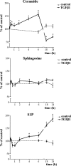
TGFβ1 affects sphingolipid metabolite levels in C2C12 myoblasts. C2C12 myoblasts were labeled for 48 h with [3-3H]sphingosine. Cellular lipids were extracted, separated, and analyzed as described in Materials and Methods section. Data represent the mean ± SEM of three independent experiments.
Figure 3.
Figure 3.
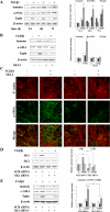
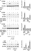
SK1 is involved in TGFβ1-dependent transdifferentiation of C2C12 myoblasts. (A) Left, C2C12 myoblasts were incubated in the absence (−) or in the presence (+) of 5 ng/ml TGFβ1 for the indicated time intervals. The content of laminin, α-SMA and transgelin (Tagln) was analyzed by Western blotting in cell lysates, using specific primary antibodies. Right, densitometric analysis. Data are reported as described in the legend to Figure 1B. (B) Left, C2C12 myoblasts were incubated in the presence of 10 μM SKI-2 60 min before 5 ng/ml TGFβ1 challenge for 24 h. Fibrosis marker proteins were detected as described in A. Right, densitometric analysis. Data are reported as described in the legend to Figure 1B. (C) C2C12 cells were seeded on microscope slides and treated as described in B. Images were obtained using a specific mouse anti-α-SMA antibody immunorevealed by a fluorescein-conjugated secondary antibody and TRITC-phalloidin staining of F-actin filaments (images are representative of three separate experiments). (D) Left, C2C12 myoblasts, transfected with scrambled (SCR) or SK1 siRNA, incubated in the absence (−) or in the presence (+) of 5 ng/ml TGFβ1 for the last 24 h of transfection, were checked for expression of SK1 or SK2 by Western blot analysis. Densitometric analysis is showed in the right panel. (E) Left, fibrosis marker proteins were detected as described in A in lysates (20 μg) of C2C12 myoblasts transfected as described in D. Right, densitometric analysis. Data are reported as described in the legend to Figure 1B.
Figure 4.
Figure 4.
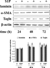
S1P does not induce transdifferentiation of C2C12 myoblasts. C2C12 myoblasts were incubated in the absence (−) or in the presence (+) of 1 μM S1P for the indicated time intervals. Top, the content of laminin, α-SMA, and transgelin (Tagln) was analyzed by Western blotting in cell lysates, using specific primary antibodies. Bottom, densitometric analysis is reported as described in the legend to Figure 1B.
Figure 5.
Figure 5.
TGFβ1 modulates the expression of S1PRs. (A) Two micrograms of total RNA extracted from C2C12 myoblasts stimulated (+) or not (−) with 5 ng/ml TGFβ1 for the indicated time intervals was subjected to semiquantitative RT-PCR analysis, using specific primers for S1P1, S1P2, S1P3, S1P4, and β-actin. (B) Quantitative mRNA analysis was performed by real-time PCR in total RNA extracted from C2C12 myoblasts stimulated (+) or not (−) with 5 ng/ml TGFβ1 for the indicated time intervals. S1PR mRNA quantitation was based on the 2−ΔΔCT method, using individual S1PR subtype of the unchallenged specimen as calibrator. (C) Quantitative mRNA analysis was performed as described in B. Results are expressed as -fold changes according to the 2−ΔΔCT method, using S1P1 as calibrator. Data are the mean ± SD of one representative experiment performed in triplicate and repeated three times with analogous results.
Figure 6.
Figure 6.
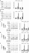
Role of S1PRs in TGFβ1-induced transdifferentiation of C2C12 myoblasts. (A) Left, confluent serum-starved C2C12 myoblasts were treated with or without 1 μM VPC23019 30 min before 5 ng/ml TGFβ1 challenge for 24 h. The content of laminin, α-SMA, and transgelin (Tagln) was analyzed by Western blotting of whole cell lysates (20 μg). Right, densitometric analysis. Data are reported as described in the legend to Figure 1B. (B) Left, confluent serum-starved C2C12 myoblasts were treated with or without 10 μM W146, 30 min before 5 ng/ml TGFβ1 challenge for 24 h. Fibrosis marker proteins were detected as described in A. Right, data are reported as described in the legend to Figure 1B. (C) C2C12 myoblasts, transfected with scrambled (SCR) or S1P3 siRNA, incubated in the absence (−) or in the presence (+) of 5 ng/ml TGFβ1 for the last 24 h of transfection, were checked for down-regulation by real-time PCR (left). Middle, fibrosis marker proteins were analyzed as described in A. Right, densitometric analysis. Data are reported as described in the legend to Figure 1B. Myoblasts transfected with SCR or S1P2 siRNA (D) and with SCR or S1P4 siRNA (E) were treated and used as described in C.
Figure 7.
Figure 7.
Dominant S1P3 signaling is required for the induction of profibrotic response by S1P. (A) Left, confluent serum-starved C2C12 myoblasts were pretreated or not with 10 μM SKI-2 for 60 min and then incubated in the presence (+) or absence (−) of 5 ng/ml TGFβ1, 6 h before the challenge with 1 μM S1P for 24 h. Transgelin (Tagln) was detected in aliquots (20 μg) of total cell lysates by Western blot analysis. Right, densitometric analysis. Data are reported as described in the legend to Figure 1B. (B) C2C12 myoblasts, transfected with scrambled (SCR) or SK1 siRNA, were incubated for 24 h in the presence (+) or absence (−) of 5 ng/ml TGFβ1, 6 h before 1 μM S1P challenge. Tagln was detected as described in A. Right, densitometric analysis. Data are reported as described in the legend to Figure 1B. (C) Left, C2C12 myoblasts, transfected with SCR or S1P3 siRNA, were treated with 10 μM SKI-2 for 60 min and then incubated in the presence (+) or absence (−) of 5 ng/ml TGFβ1, 6 h before 1 μM S1P challenge. Tagln was detected as described in A. Right, densitometric analysis. Data are reported as described in the legend to Figure 1B. (D) Left, C2C12 cells were transiently transfected with pcDNA3-S1P3 or empty vector. Cells were treated with 1 μM S1P or 5 ng/ml TGFβ1 for 24 h and cell lysates used to detect Tagln as described in A. Inset, S1P3 expression levels was evaluated in transfected C2C12 myoblasts by Western blot analysis using specific monoclonal anti-HA antibodies. Right, densitometric analysis. Data are reported as described in the legend to Figure 1B.
Figure 8.
Figure 8.
Role of Smad proteins in TGFβ1-dependent SK1 and S1P3 up-regulation and in TGFβ1-induced transdifferentiation of C2C12 myoblasts. C2C12 myoblasts were transfected with Smad2 or Smad3 or Smad4 siRNA, using scrambled (SCR) siRNA as a control. (A) Top, C2C12 myoblasts were checked for expression of Smad2, Smad3, or Smad4 by Western blot analysis, using specific anti-Smad2/3 or anti-Smad4 antibodies. Densitometric analysis is showed in the bottom panels. Data are reported as described in the legend to Figure 1B. (B) Left, C2C12 myoblasts, transfected with SCR, Smad2, Smad3, or Smad4 siRNA, were incubated in the presence (+) or absence (−) of 5 ng/ml TGFβ1 for 24 h. Total cell lysates (20 μg) were subjected to Western blot analysis to detect SK1 and Transgelin (Tagln) expression. Right, densitometric analysis is reported as described in the legend to Figure 1B. (C) Quantitative analysis of S1P3 mRNA was performed by real-time PCR in total RNA extracted from C2C12 myoblasts transfected with SCR, Smad2, Smad3, or Smad4 siRNA, stimulated (+) or not (−) with 5 ng/ml TGFβ1 for 24 h. Results are expressed as -fold changes according to the 2−ΔΔCT method, using S1P3 subtype of the unchallenged specimen as calibrator. Data are the mean ± SD of one representative experiment performed in triplicate and repeated three times with analogous results.
Figure 9.
Figure 9.
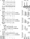
TGFβ1-dependent transdifferentiation of C2C12 myoblasts is dependent on RhoA/Rho kinase activation, downstream of SK1 and S1P3. (A) Left, C2C12 myoblasts were treated with or without 10 μM U0126 or 25 μM Y27632, 30 min before 5 ng/ml TGFβ1 challenge for 24 h. The content of SK1 and transgelin (Tagln) was analyzed by Western analysis of whole cell lysates (20 μg). Right, densitometric analysis. A blot representative of three independent experiments is shown. Data are reported as described in the legend to Figure 1B. (B) Left, C2C12 myoblasts were incubated for the indicated time intervals with or without 5 ng/ml TGFβ1 in the presence or absence of 10 μM SKI-2. Western analysis of RhoA was performed in membrane and cytosolic fractions (10 μg). Right, densitometric analysis of three independent experiments. Data are reported as described in the legend to Figure 1D. (C) Left, C2C12 myoblasts were treated with or without 5 ng/ml TGFβ1 for 8 h in the presence or in the absence of 1 μM VPC23019. Western analysis was performed as described in B. Right, densitometric analysis. Data are reported as described in the legend to Figure 1D. (D) C2C12 myoblasts, transfected with scrambled (SCR), SK1, or S1P3 siRNA, were incubated in the presence (+) or absence (−) of 5 ng/ml TGFβ1 for 8 h. Left, Western analysis was performed as described in B. Right, densitometric analysis is reported as described in the legend to Figure 1D.
Figure 10.
Figure 10.
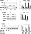
SK1/S1P3 pathway is implicated in the antimyogenic action of TGFβ1. Top left, C2C12 myoblasts were incubated for the indicated time-intervals with (+) or without (−) 5 ng/ml TGFβ1. The content of MHC and caveolin-3 (Cav 3) was analyzed by Western blot in aliquots (20 μg) of whole cell lysates. Top right, densitometric analysis. Data are reported as described in the legend to Figure 1B. Middle left, C2C12 myoblasts were incubated in the presence of 10 μM SKI-2, 60 min before 5 ng/ml TGFβ1 challenge for 48 h and analyzed for myogenic marker expression. Middle right, densitometric analysis. Data are reported as described in the legend to Figure 1B. Bottom left, C2C12 myoblasts, transfected with scrambled (SCR), SK1, or S1P3 siRNA, were incubated in the presence (+) or absence (−) of 5 ng/ml TGFβ1. The content of Cav 3 was analyzed as described in A. Bottom right, densitometric analysis is reported as described in the legend to Figure 1B.
Figure 11.
Figure 11.
Working model of the mechanism by which TGFβ1 elicits myoblast transdifferentiation into myofibroblasts.

References

    1. Alvarez S. E., Milstien S., Spiegel S. Autocrine and paracrine roles of sphingosine-1-phosphate. Trends Endocrinol. Metab. 2007;18:300–307. - PubMed
    1. Best T. M., Hunter K. D. Muscle injury and repair. Phys. Med. Rehabil. Clin. N. Am. 2000;11:251–266. - PubMed
    1. Cohn R. D., et al. Angiotensin II type 1 receptor blockade attenuates TGF-beta-induced failure of muscle regeneration in multiple myopathic states. Nat. Med. 2007;13:204–210. - PMC - PubMed
    1. Derynck R., Zhang Y. E. Smad-dependent and Smad-independent pathways in TGF-beta family signalling. Nature. 2003;425:577–584. - PubMed
    1. Dolman M. E., Fretz M. M., Segers G. J., Lacombe M., Prakash J., Storm G., Hennink W. E., Kok R. J. Renal targeting of kinase inhibitors. Int. J. Pharm. 2008;364:249–257. - PubMed

Publication types

MeSH terms

LinkOut - more resources