. 2010 Mar 15;21(6):970-8.
doi: 10.1091/mbc.e09-07-0638. Epub 2010 Jan 20.

Differential localization and dynamics of class I myosins in the enterocyte microvillus

Affiliations

Differential localization and dynamics of class I myosins in the enterocyte microvillus

Andrew E Benesh et al. Mol Biol Cell. .

Abstract

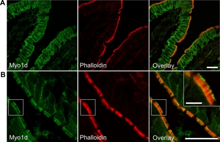
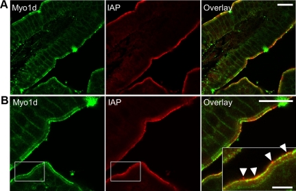
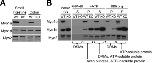
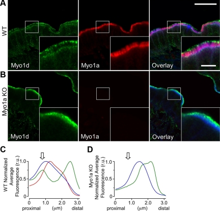
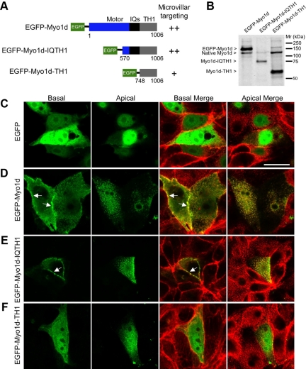
Epithelial cells lining the intestinal tract build an apical array of microvilli known as the brush border. Each microvillus is a cylindrical membrane protrusion that is linked to a supporting actin bundle by myosin-1a (Myo1a). Mice lacking Myo1a demonstrate no overt physiological symptoms, suggesting that other myosins may compensate for the loss of Myo1a in these animals. To investigate changes in the microvillar myosin population that may limit the Myo1a KO phenotype, we performed proteomic analysis on WT and Myo1a KO brush borders. These studies revealed that WT brush borders also contain the short-tailed class I myosin, myosin-1d (Myo1d). Myo1d localizes to the terminal web and striking puncta at the tips of microvilli. In the absence of Myo1a, Myo1d peptide counts increase twofold; this motor also redistributes along the length of microvilli, into compartments normally occupied by Myo1a. FRAP studies demonstrate that Myo1a is less dynamic than Myo1d, providing a mechanistic explanation for the observed differential localization. These data suggest that Myo1d may be the primary compensating class I myosin in the Myo1a KO model; they also suggest that dynamics govern the localization and function of different yet closely related myosins that target common actin structures.


Figures

Figure 1.
Figure 1.
Myo1d localizes to the enterocyte basolateral membrane, terminal web, and microvillar tips. Representative frozen sections of mouse small intestine stained with antibodies targeting Myo1d (green) and Alexa 488 (or 633)–conjugated phalloidin (red). (A and B) Myo1d C13 antibody targets the basolateral membrane, terminal web, and microvillar tips of enterocytes. Bar, 20 μm; inset bar, 5 μm.
Figure 2.
Figure 2.
Myo1d and IAP partially colocalize at tips of microvilli. Frozen sections of mouse small intestine labeled with antibodies targeting Myo1d (green) and IAP (red). (A) Confocal image of a villus demonstrating both Myo1d and IAP localize to microvillar tips. (B) Representative view of partial colocalization between Myo1d and IAP at microvillar tips. Myo1d at microvillar tips appears in two populations: alone in distinct puncta, and colocalized with IAP. Arrowheads mark IAP-enriched puncta that lack Myo1d at the extreme distal tips of microvilli. Bars, (A and B) 20 μm; inset in B, 5 μm.
Figure 3.
Figure 3.
Myo1d redistributes within the enterocyte in the absence of Myo1a. (A) Whole cell lysates were created from WT and KO small intestine and colon mucosal scrapings and then blotted for Myo1a and Myo1d. (B) Compartmentalization of Myo1d within the BB is Myo1a-dependent. Myo1d protein levels are slightly higher in KO BBs compared with WT BBs. Neither Myo1d nor Myo1a are found with detergent-solubilized membranes (DSMs) from whole BBs after treatment with 1% NP-40 (+NP-40, S). On treatment with mM ATP, Myo1a distributes equally between soluble (+ATP, S) and insoluble (+ATP, P) fractions. Most Myo1d is found in the supernatant, with low levels remaining in the pellet. In the absence of Myo1a, the amount of Myo1d associated with actin bundles increases dramatically. Centrifugation of the ATP supernatant (+ATP, S) at 100K × g enables the separation of DRMs (100K × g, P) from purely soluble proteins (100K × g, S). In WT samples, Myo1a partitions equally between the DRM and soluble fractions, whereas Myo1d does not sediment with DRMs. However, Myo1d signal appears in the DRM-enriched 100K × g pellet in Myo1a KO samples. The 100K × g gel samples were concentrated 2.5-fold relative to all other samples. Because Myo2 is a BB component that is not expected to change in the absence of Myo1a (see Table 1), blots for Myo2 are shown as a loading control.
Figure 4.
Figure 4.
Myo1a and Myo1d exhibit differential localization within the BB. (A) Confocal images of adult WT mouse small intestine frozen sections stained for Myo1d (green, C13 antibody), Myo1a (red), and F-actin (blue). In the BB, Myo1d occupies microvillar tips and the terminal web, whereas Myo1a localizes along the length of microvilli. (B) KO mouse sections stained in an identical manner reveal that Myo1d is found along the length of microvilli in the absence of Myo1a. Myo1d still occupies microvillar tips, but redistributes from lateral plasma membrane and the terminal web. (C and D) Plots show the average pixel intensity along the microvillar axis from proximal (base) to distal (tip) for Myo1d (green), Myo1a (red), and phalloidin (blue) fluorescence signals. The arrow indicates the position of the terminal web. Representative micrographs of “straightened” BBs used to create these plots are shown in Supplemental Figure 3. Bar, 20 μm; inset bar, 5 μm; bars serve as calibration for A and B.
Figure 5.
Figure 5.
Truncation analysis reveals that both IQ and TH1 domains are needed for proper localization of Myo1d. (A) Diagram of constructs that were generated for studying Myo1d localization determinants. EGFP was fused to the N-terminus of the full-length molecule, Myo1d-IQTH1, or Myo1d-TH1. An indication of the ability of each construct to target to microvilli is provided to the right of the diagram. (B) Western blots with the H60 anti-Myo1d antibody confirm that fragments of the expected size are produced in CL4 cells. (C–F) Confocal micrographs of CL4 cells expressing one of three EGFP-tagged Myo1d constructs or EGFP alone (green) and counterstained with Alexa 488– or Alexa 633–conjugated phalloidin (red). Merges of green and red channels are shown at both apical and basal planes. (C) EGFP alone demonstrates diffuse localization throughout the cytosol and nucleus. (D) EGFP-Myo1d is enriched in microvilli and also targets the lateral plasma membrane, in a manner similar to that previously reported for EGFP-Myo1a (Tyska and Mooseker, 2002). (E) EGFP-Myo1d-IQTH1 localizes to microvilli and lateral membranes in a manner similar to full-length Myo1d. (F) EGFP-Myo1d-TH1 demonstrates low level targeting to microvilli and plasma membrane. Bar, (A) 20 μm (serves as a calibration for A–E).
Figure 6.
Figure 6.
Myo1a and Myo1d demonstrate differential dynamics in the BB. (A) EGFP-Myo1d and (B) EGFP-Myo1a were expressed in CL4 cells and grown on filters for FRAP studies. Micrographs show representative examples of photobleaching; ROIs are marked with an arrow. (C) Averaged datasets (n = 14 for EGFP-Myo1a, n = 22 for EGFP-Myo1d) of the relative fluorescence recovery in photobleached regions were fit to a general kinetic model as outlined in Methods. Myo1d (green) demonstrates more complete recovery (i.e., a higher mobile fraction) when compared with Myo1a (red). (D) Stacked bar graphs of the amplitudes for Myo1d and Myo1a (Fast phase, black; Slow phase, gray). (E) Bar graphs of the rate constants for Myo1d and Myo1a (Fast phase, black; Slow phase, gray). Bar, (A) 20 μm (serves as a calibration for A and B).

Similar articles

Cited by

References

    1. Bahler M., Kroschewski R., Stoffler H. E., Behrmann T. Rat myr 4 defines a novel subclass of myosin I: identification, distribution, localization, and mapping of calmodulin-binding sites with differential calcium sensitivity. J. Cell Biol. 1994;126:375–389. - PMC - PubMed
    1. Belyantseva I. A., Boger E. T., Friedman T. B. Myosin XVa localizes to the tips of inner ear sensory cell stereocilia and is essential for staircase formation of the hair bundle. Proc. Natl. Acad. Sci. USA. 2003;100:13958–13963. - PMC - PubMed
    1. Belyantseva I. A., Boger E. T., Naz S., Frolenkov G. I., Sellers J. R., Ahmed Z. M., Griffith A. J., Friedman T. B. Myosin-XVa is required for tip localization of whirlin and differential elongation of hair-cell stereocilia. Nat. Cell Biol. 2005;7:148–156. - PubMed
    1. Berg J. S., Cheney R. E. Myosin-X is an unconventional myosin that undergoes intrafilopodial motility. Nat. Cell Biol. 2002;4:246–250. - PubMed
    1. Cao Z., et al. Use of fluorescence-activated vesicle sorting for isolation of Naked2-associated, basolaterally targeted exocytic vesicles for proteomics analysis. Mol. Cell Proteom. 2008;7:1651–1667. - PMC - PubMed

Publication types