. 2010 Mar 15;21(6):897-904.
doi: 10.1091/mbc.e09-07-0598. Epub 2010 Jan 20.

A specific form of phospho protein phosphatase 2 regulates anaphase-promoting complex/cyclosome association with spindle poles

Affiliations

A specific form of phospho protein phosphatase 2 regulates anaphase-promoting complex/cyclosome association with spindle poles

Jorge Z Torres et al. Mol Biol Cell. .

Abstract

In early mitosis, the END (Emi1/NuMA/Dynein-dynactin) network anchors the anaphase-promoting complex/cyclosome (APC/C) to the mitotic spindle and poles. Spindle anchoring restricts APC/C activity, thereby limiting the destruction of spindle-associated cyclin B and ensuring maintenance of spindle integrity. Emi1 binds directly to hypophosphorylated APC/C, linking the APC/C to the spindle via NuMA. However, whether the phosphorylation state of the APC/C is important for its association with the spindle and what kinases and phosphatases are necessary for regulating this event remain unknown. Here, we describe the regulation of APC/C-mitotic spindle pole association by phosphorylation. We find that only hypophosphorylated APC/C associates with microtubule asters, suggesting that phosphatases are important. Indeed, a specific form of PPP2 (CA/R1A/R2B) binds APC/C, and PPP2 activity is necessary for Cdc27 dephosphorylation. Screening by RNA interference, we find that inactivation of CA, R1A, or R2B leads to delocalization of APC/C from spindle poles, early mitotic spindle defects, a failure to congress chromosomes, and decreased levels of cyclin B on the spindle. Consistently, inhibition of cyclin B/Cdk1 activity increased APC/C binding to microtubules. Thus, cyclin B/Cdk1 and PPP2 regulate the dynamic association of APC/C with spindle poles in early mitosis, a step necessary for proper spindle formation.


Figures

Figure 1.
Figure 1.
Depletion of the PPP2 subunits CA, R1A, or R2B leads to defective spindle formation and chromosome congression. (A) HeLa cells were transfected with siRNA targeting CA, R1A, or R2B for 48 h. The extent of gene knockdown was assessed by immunoblotting with antibodies directed against CA, R1A, R2B, and actin or GAPDH as loading controls. (B) Depletion of CA, R1A, or R2B leads to centrosome, mitotic spindle assembly, and chromosome congression defects. RNAi-treated cells in A were fixed and stained with Hoechst 33342, anti-α-tubulin, anti-pericentrin, and anti-centrin antibodies to visualize DNA, the mitotic spindle, the pericentriolar material, and the centrioles, respectively. Three groups of cell phenotypes were observed upon depletion of PPP2 subunits and are designated as groups 1 through 3. Group 1, has centrosome amplification and chromosome congression defects along with multipolar spindle formation. Group 2, contains chromosome cohesion defects and defective spindles. Group 3, exhibits defective spindle formation, spindle pole fragmentation, and a failure to congress chromosomes properly. Bar, 5 μm.
Figure 2.
Figure 2.
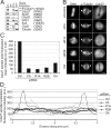
Association of Cdc27 with spindle poles requires the PPP2 subunits CA, R1A, and R2B. (A) Dephosphorylated APC/C preferentially associates with microtubule asters in vitro. Mitotic HeLa cell extracts were used for in vitro microtubule polymerization reactions in the presence or absence of taxol. Polymerized microtubules were subjected to centrifugation, and samples from the supernatant (S) and pelleted (P) microtubules were analyzed by immunoblot. Immunoblotting was used to detect the association of the APC/C subunits Cdc27, Apc8, Cdc20, Cdh1, and positive (mitotic kinesin Eg5) and negative (CycD) controls with microtubules. The upper and lower arrows indicate phosphorylated and dephosphorylated forms of Cdc27, respectively. Tubulin polymerization was confirmed by staining the membrane with Coomassie blue. (B) Depletion of CA, R1A, or R2B by siRNA leads to delocalization of Cdc27 from spindle poles. RNAi-treated cells (as in Figure 1) or cells treated acutely with 175 nM OA for 13 min were fixed and stained with Hoechst 33342 and anti-α-tubulin and anti-Cdc27 antibodies to visualize DNA, the mitotic spindle, and Cdc27, respectively. Bar, 5 μm. (C) Quantitation of mean intensity of Cdc27 at spindle poles. A 2-μm square was drawn around each of 20 spindle poles from control, CA, R1A, or R2B siRNA-treated cells or cells treated acutely with OA, and the mean fluorescence intensity of Cdc27 spindle pole staining was quantified and presented as arbitrary units (AU). (D) Spindle associated Cdc27 is reduced in mitotic cells depleted of CA, R1A, or R2B or treated acutely with OA. Line intensity measurements taken along axis intersecting the two poles (marked by arrows in A) were quantified and graphed. X-axis, distance in micrometers from the center of the two poles. Y-axis, fluorescence intensities along the axis in arbitrary units (AU).
Figure 3.
Figure 3.
Dynamic association of the APC/C with mitotic microtubule asters is regulated by a kinase-phosphatase equilibrium. (A) Inhibition of Cdk activity and an OA-sensitive phosphatase in vivo perturbs the association of APC/C with microtubules. Mitotic HeLa cells were treated with control vehicle, the Cdk inhibitor roscovitine (Rosco; 1 h), or the phosphatase inhibitor okadaic acid (OA; 30 min) before extract preparation. Mitotic microtubule copelleting reactions (as in Figure 2) were performed in the presence of control vehicle, Rosco, or OA. Supernatant and pellet fractions were immunoblotted with antibodies against APC/C subunits/coactivators (Cdc27, Apc8, Cdc20, and Cdh1) or Eg5 to detect microtubule association. Cdc27 phosphorylation status, association with microtubules, and tubulin polymerization was monitored, as described in Figure 2B. For A and B, arrows indicate phosphorylated and dephosphorylated forms of Cdc27. (B) Phosphorylation and dephosphorylation of APC/C in vitro perturbs APC/C microtubule association. Mitotic HeLa cells were treated with control vehicle, Rosco, or OA as described in A before extract preparation. Extracts from Rosco-treated cells were incubated with cyclin B/Cdk1 for 20 min at 30°C. Conversely, extracts from OA-treated cells were incubated with lambda phosphatase (λPPase) for 20 min at 30°C. All extracts were subjected to mitotic microtubule copelleting reactions and the status of Cdc27 phosphorylation, its association with microtubules, and tubulin polymerization was detected as described in Figure 2B.
Figure 4.
Figure 4.
PPP2 associates with mitotic microtubules, binds Cdc27, and is required for Cdc27 dephosphorylation. (A) PPP2 associates with microtubules. Mitotic HeLa cell extracts were used for in vitro microtubule polymerization reactions in the presence or absence of taxol. Polymerized microtubules were subjected to centrifugation and samples from the supernatant (S) and pelleted (P) microtubules were analyzed by Western blot. Antibodies directed against the catalytic subunit of OA-sensitive phosphatases (PPP1–PPP6) and the PPP2 regulatory subunits R1A and R2B were used to detect their association with microtubules. Tubulin polymerization was detected using rat anti-α-tubulin antibody. (B) Emi1 is efficiently immunodepleted from HeLa cell extracts. G1/S (Interphase) or G2/M (Mitosis) HeLa cell extracts, 500 μg, were incubated with 10 μg of control IgG (Ig) or anti-Emi1 antibodies. Depleted extracts were probed with anti-Emi1 and GAPDH antibodies. (C) Cdc27 associates with PPP2. G1/S and G2/M extracts depleted of Emi1, from B, were incubated with 10 μg of control IgG or anti-Cdc27 antibodies. Immunoprecipitations were probed with anti-Cdc27, -Cdh1, -PPP1CA, -PPP2CA, -PPP2R2B, and -GAPDH antibodies. IgGlc indicates the IgG light chain cross-reacting band. (D) PPP2 knockdown inhibits Cdc27 dephosphorylation. Mitotic HeLa cell extracts were prepared from siRNA-treated cells targeting the catalytic subunit of each member of the family of OA-sensitive phosphatases. Extracts were incubated at 37°C and samples taken at 0-, 30-, and 60-min time points. The extent of Cdc27 dephosphorylation was monitored by immunoblot using anti-Cdc27 antibodies. The upper and lower arrows indicate phosphorylated and dephosphorylated forms of Cdc27, respectively.
Figure 5.
Figure 5.
PPP2 activity is required to maintain proper cyclin B levels on the spindle. (A) Knockdown of PPP2 subunits, (CA, R1A, or R2B), reduces pole-associated cyclin B levels. HeLa cells were transfected with control, CA, R1A, or R2B siRNA for 48 h before fixation. Staining with Hoechst 33342, anti-α-tubulin, and anti-cyclin B visualized DNA, the mitotic spindle, and cyclin B localization, respectively. Bar, 5 μm. (B) Quantitation of mean fluorescence intensity of cyclin B at spindle poles as described in Figure 2C for control, CA, R1A, and R2B siRNA-treated cells. (C) Spindle associated cyclin B is reduced in mitotic cells depleted of CA, R1A, or R2B. Line intensity measurements taken along an axis intersecting the two poles (marked by arrows in A) were quantified and graphed. X-axis, distance, in micrometers, from the center of the two poles. Y-axis, fluorescence intensities along the axis in arbitrary units (AU).
Figure 6.
Figure 6.
Model for APC/C association with mitotic spindle poles. Mitotic kinases and phosphatases control the dynamic association of the APC/C with mitotic spindle poles. The PPP2 (CA/R1A/R2B) phosphatase is necessary for APC/C dephosphorylation and regulates its association with the minus ends of microtubules (spindle poles), possibly via the END network. Local inhibition of APC/C activity at spindle poles in early mitosis allows for stabilization of cyclin B/Cdk1 activity near poles, an activity necessary for mitotic spindle formation. Phosphorylation of the APC/C by cyclin B/Cdk1 inhibits its association with spindle poles. Perturbation of this equilibrium alters APC/C localization and results in mitotic spindle defects and a failure to congress chromosomes.

Similar articles

Cited by

References

    1. Andersen J. S., Wilkinson C. J., Mayor T., Mortensen P., Nigg E. A., Mann M. Proteomic characterization of the human centrosome by protein correlation profiling. Nature. 2003;426:570–574. - PubMed
    1. Arachchige Don A. S., Dallapiazza R. F., Bennin D. A., Brake T., Cowan C. E., Horne M. C. Cyclin G2 is a centrosome-associated nucleocytoplasmic shuttling protein that influences microtubule stability and induces a p53-dependent cell cycle arrest. Exp. Cell Res. 2006;312:4181–4204. - PMC - PubMed
    1. Ban K. H., Torres J. Z., Miller J. J., Mikhailov A., Nachury M. V., Tung J. J., Rieder C. L., Jackson P. K. The END network couples spindle pole assembly to inhibition of the anaphase-promoting complex/cyclosome in early mitosis. Dev. Cell. 2007;13:29–42. - PubMed
    1. Belmont L. D., Hyman A. A., Sawin K. E., Mitchison T. J. Real-time visualization of cell cycle-dependent changes in microtubule dynamics in cytoplasmic extracts. Cell. 1990;62:579–589. - PubMed
    1. Chen F., et al. Multiple protein phosphatases are required for mitosis in Drosophila. Curr. Biol. 2007;17:293–303. - PubMed

Publication types