. 2010 Mar 15;21(6):860-70.
doi: 10.1091/mbc.e09-07-0567. Epub 2010 Jan 20.

The RhoA activator GEF-H1/Lfc is a transforming growth factor-beta target gene and effector that regulates alpha-smooth muscle actin expression and cell migration

Affiliations

The RhoA activator GEF-H1/Lfc is a transforming growth factor-beta target gene and effector that regulates alpha-smooth muscle actin expression and cell migration

Anna Tsapara et al. Mol Biol Cell. .

Abstract

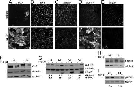
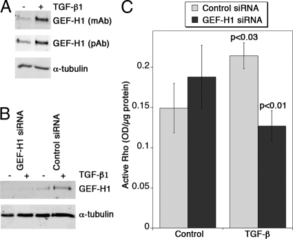
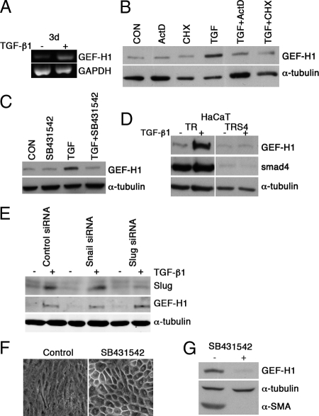
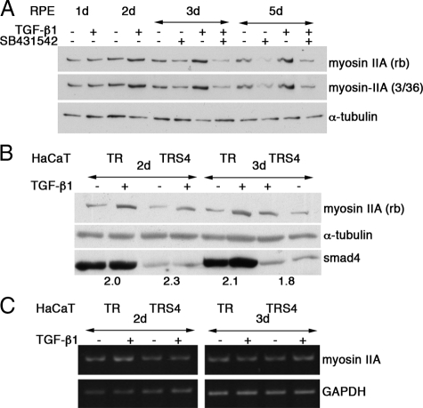
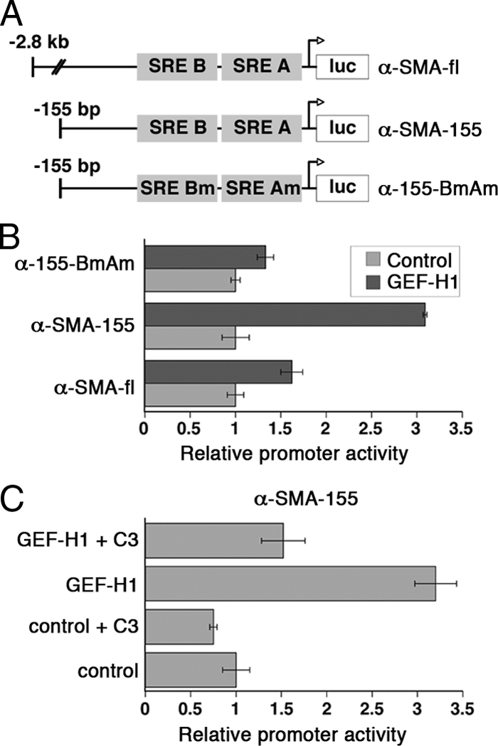
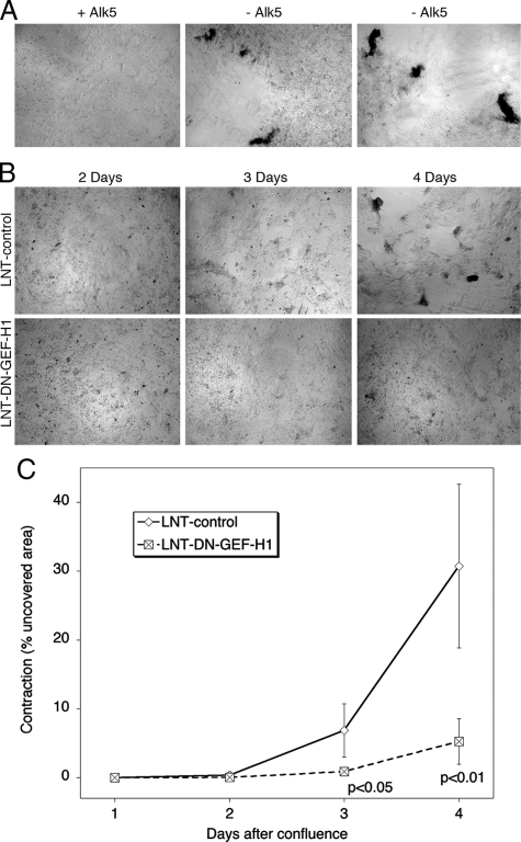
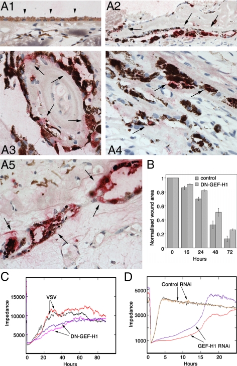
Maintenance of the epithelial phenotype is crucial for tissue homeostasis. In the retina, dedifferentiation and loss of integrity of the retinal pigment epithelium (RPE) leads to retinal dysfunction and fibrosis. Transforming growth factor (TGF)-beta critically contributes to RPE dedifferentiation and induces various responses, including increased Rho signaling, up-regulation of alpha-smooth muscle actin (SMA), and cell migration and dedifferentiation. Cellular TGF-beta responses are stimulated by different signal transduction pathways: some are Smad dependent and others Smad independent. Alterations in Rho signaling are crucial to both types of TGF-beta signaling, but how TGF-beta-stimulates Rho signaling is poorly understood. Here, we show that primary RPE cells up-regulated GEF-H1 in response to TGF-beta. GEF-H1 was the only detectable Rho exchange factor increased by TGF-beta1 in a genome-wide expression analysis. GEF-H1 induction was Smad4-dependant and led to Rho activation. GEF-H1 inhibition counteracted alpha-SMA up-regulation and cell migration. In patients with retinal detachments and fibrosis, migratory RPE cells exhibited increased GEF-H1 expression, indicating that induction occurs in diseased RPE in vivo. Our data indicate that GEF-H1 is a target and functional effector of TGF-beta by orchestrating Rho signaling to regulate gene expression and cell migration, suggesting that it represents a new marker and possible therapeutic target for degenerative and fibrotic diseases.


Figures

Figure 1.
Figure 1.
TGF-β1 induces junctional disruption and GEF-H1 up-regulation in RPE cells. RPE cells were stimulated with TGF-β1 (A–E for 3 d; F–I as indicated) and processed for immunofluorescence (A–E) or immunoblot (F–H) analysis. (A–C) Samples were stained for either α-SMA (A) and ZO-1 (B), occludin (C), or GEF-H1 (D) and cingulin (E). (F–H) Immunoblots of total RPE cell extracts stimulated with TGF-β1 for the indicated time were probed with antibodies against ZO-1 and occludin (by densitometry, both proteins were decreased by >50% after 3 and 5 d of TGF-β treatment; F), GEF-H1 and α-SMA (the numbers indicate the ratios of TGF-β–treated divided by control samples obtained by densitometry; all values were normalized by those obtained for tubulin in each sample; G), cingulin (H); α-tubulin was used as loading control. (I) Immunoblot of RPE cell extracts was probed for phosphorylated (p-MYPT1) and total myosin light chain phosphatase (MYPT1) (the numbers indicate the relative increase in p-MYPT1 in TGF-β–treated samples). Shown are representative results from at least two experiments.
Figure 2.
Figure 2.
Inhibition of TGF-β–induced Rho activity by GEF-H1 depletion. (A) HaCaT cells were stimulated for 3 d with TGF-β and were then analyzed for expression of GEF-H1 and α-tubulin. Expression of GEF-H1 was monitored with two different antibodies, an mAb antibody and a polyclonal (pAb) antibody that recognize different epitopes. (B and C) HaCaT cells were transfected with control or GEF-H1 targeting siRNAs, and, after 24 h, were incubated with or without TGF-β for the next 3 d. The cells were then lysed to monitor expression of GEF-H1 (B) or analyzed for active RhoA levels (C; shown are means ± 1 SD, n = 3; indicated are p values obtained from t tests comparing TGF-β-treated cells with not-treated control siRNA-transfected cells and, respectively, GEF-H1 depleted TGF-β–treated cells with control siRNA transfected TGF-β–treated cells).
Figure 3.
Figure 3.
TGF-β1-induced GEF-H1 up-regulation is Smad4 dependent. (A) RT-PCR analysis for GEF-H1 mRNA levels in control and TGF-β1–treated (3-d) samples; GAPDH was used as a loading control. Note that the increase observed by RT-PCR (>2-fold) was similar to the increase obtained from the microarray analysis. (B) RPE cells were preincubated with actinomycin D (ActD) or cycloheximide (CHX) and then stimulated or not with TGF-β1 for 2 d. Immunoblot of total RPE cell extracts probed for GEF-H1 and α-tubulin. Note that the TGF-β–induced increase was blocked by >85% by both actinomycin D and cycloheximide. (C) RPE cells were preincubated with SB431542, TGF-β receptor type I kinase inhibitor, and then stimulated with TGF-β1 for 3 d and tested for GEF-H1 and α-tubulin expression. (D) HaCaT-TR-S4, a stable cell line permitting inducible depletion of Smad4, and the parental cell line HaCaT-TR were treated with tetracycline for 2 d to reduce Smad4 expression and were then stimulated with TGF-β1 for 4 d. Total cell extracts were probed for GEF-H1 and Smad4. Note: A threefold up-regulation of GEF-H1 was observed in HaCaT cells that was blocked by Smad4 depletion. (E) HaCaT cells were transfected with control or Slug-targeting siRNAs. After 24 h, the cells were incubated with fresh medium without or with TGF-β for 3 d before analysis of Slug, GEF-H1 and tubulin expression. Note that no change in GEF-H1 expression was observed upon depletion of Slug. (F and G) RPE cells plated at very low density were grown in the absence or presence of the ALK5 inhibitor SB431542 for 14 d. (E) Phase contrast images of control and treated cells. (G) Immunoblot for GEF-H1, α-tubulin, and α-SMA.
Figure 4.
Figure 4.
TGF-β1–induced myosin-IIA up-regulation is Smad4 independent. (A) RPE cultures in the absence or presence of the ALK45 kinase inhibitor SB431542 were stimulated TGF-β1 as indicated. Immunoblots of total cell extracts are shown that were probed sequentially for myosin-IIA by using two different antibodies, a rabbit antibody (Sigma-Aldrich) or monoclonal (mouse, 3/36); α-tubulin was used as loading control. By densitometry, myosin-II was up-regulated by at least 55%. (B) HaCaT-TR-S4, a stable clone for inducible depletion of Smad4, and the parental cell line HaCaT-TR were treated with tetracycline for 2 d to reduce Smad4 expression and then stimulated with TGF-β1 for the indicated times. Total cell extracts were probed for myosin-IIA (rabbit; Sigma-Aldrich) and Smad4. The numbers indicate the ratio between TGF-β–treated and control samples for myosin-II. (C) RT-PCR analysis for myosin-IIA in control and TGF-β1–treated HaCaT-TR and HaCaT-TR-S4 cells; GAPDH served as a control to monitor RNA input. By densitometry, no significant differences were observed between control and TGF-β–treated samples.
Figure 5.
Figure 5.
GEF-H1 regulates the α-SMA promoter in a Rho-dependent manner. (A) Schematic representation of the α-SMA promoter constructs used. α-SMA-fl is a 2.8-kb full-length α-SMA promoter; α-SMA-155 is a minimal promoter (155-base pairs) containing the two SREs and lacking upstream inhibitory elements; and α-155-BmAm is an identical minimal region promoter in which the SREs had been mutated. The relative position of the two SREs (SRE A and SRE B) is indicated. (B) RPE cells were cotransfected with a α-SMA firefly luciferase reporter construct and a control Renilla luciferase promoter plasmid together with either expression (GEF-H1) or empty (control) vector. The results are expressed as percentage of control transfections (shown are means ± 1 SD of 4 determinations). Note that the full-length promoter contains additional upstream regulatory elements that suppress full activation in response to Rho stimulation. (C) The α-SMA-155 reporter construct was used and cells were incubated with TAT-C3, a membrane-permeable C3 transferase.
Figure 6.
Figure 6.
Rho signaling and GEF-H1 regulate α-SMA expression induced by TGF-β1. (A) RPE cells were incubated for 3 d with or without TGF-β1. During the last 2 d, membrane-permeable C3 transferase was added as indicated. Expression of GEF-H1 and α-tubulin was then analyzed by immunoblotting. (B and C) RPE cells were transiently transfected with DN-GEF-H1 and treated with TGF-β1 for 3 d. The cells were then fixed and processed for immunofluorescence using antibodies against α-SMA and VSV, to detect DN-GEF-H1. Shown is an example of obtained images (B), and quantifications of percentages of α-SMA–positive cells in the control (VSV-negative) and DN-GEF-H1 expressing (VSV- positive) cell populations. (D) RPE cells were infected with control (LNT-control) or DN-GEF-H1 (LNT-DN-GEF-H1) lentivirus and stimulated with TGF-β1 for 3 d, and then α-SMA and fibronectin expression was analyzed in total cell extracts. The graphs show densitometric analysis of scanned immunoblot data. Note: Dominant-negative GEF-H1 inhibits TGF-β1–induced α-SMA expression.
Figure 7.
Figure 7.
GEF-H1 inhibition counteracts TGF-β–dependent monolayer contraction and detachment. (A) Three-week-old primary cultures of porcine RPE cells were plated at high density (6 × 10⁴ cells/cm2) on fibronectin-coated coverslips. The confluent monolayers were then incubated with or without the ALK5 inhibitor SB431542. Note, monolayers in which TGF-β signaling was not inhibited contracted and started to detach from the substrate. (B and C) Three-week-old primary cultures of porcine RPE cells infected with a control lentivirus (LNT-control) or a virus encoding dominant negative GEF-H1 (LNT-DN-GEF-H1) were cultured as in panel A without the ALK5 inhibitor and inspected daily. Cell-free areas were then quantified and expressed as percentage of total area. The quantification in C is based on the analysis of four-independent cultures per condition. Indicated are p values derived from a t test.
Figure 8.
Figure 8.
GEF-H1 is up-regulated in migratory RPE cells in vivo and regulates cell migration. (A1) Negative GEF-H1 staining was observed in RPE cells from a normal-looking area adjacent to a choroidal malignant melanoma (case 1, 40×). (A2) Positive GEF-H1 staining in elongated migratory RPE configured as a monolayer (case 2, trauma, 40×). (A3) Positive GEF-H1 staining in migratory RPE around vessels from a case of posterior Uveitis (case 3, 40×). (A4) Positive GEF-H1 staining in migratory pigmented RPE within an area of subretinal scaring from a case of corneal infection (case 4, 40×). (A5) Positive GEF-H1 staining in apex to apex islands of RPE cells from a case of retinal detachment (case 5, 40×). In all instances. a red chromogen was used and sections were counterstained with hematoxylin. Note that RPE cells can be recognized by their strong pigmentation and are classified as migratory when they are displaced form their normal location at the back of the retina and have moved into the neural retina. In A1, the RPE is indicated with arrow heads, and the arrows in A2–A5 point to groups of RPE cells positive for GEF-H1. (B) RPE cells infected with LNT-control or LNT-DN-GEF-H1 were treated with TGF-β1 for 3 d, and then a wound was manually inflicted with a pipette tip. Pictures were then taken after 16, 24, 48, and 72 h, and the wound area was quantified. The wound areas were normalized to the areas obtained at 0 h that are referred as 1, and all other areas were then expressed as fractions of the initial wound, the graphs represent normalized wound areas at different times (shown are averages ± 1 SD; n = 4). (C) RPE cells infected with LNT-control or LNT-DN-GEF-H1 were treated with TGF-β1 for 3 d and then subjected to a high electric field to induce a wound in the center of each monolayer. The graphs represent wound closure as measured by recovery of impedance along time (2 separate measurements for each condition are shown that had been analyzed in parallel and correspond to a representative experiment). (D) MDCK cells, control cells or cells permitting tetracycline-induced GEF-H1 depletion by RNA interference (Benais-Pont et al., 2003), were cultured with the antibiotic and then subjected to a high electric field to induce a wound in the center of each monolayer and impedance was measured to monitor wound closure. Shown is a representative experiment performed in duplicates.

Similar articles

Cited by

References

    1. Ablonczy Z., Crosson C. E. VEGF modulation of retinal pigment epithelium resistance. Exp. Eye Res. 2007;85:762–771. - PMC - PubMed
    1. Aijaz S., D'Atri F., Citi S., Balda M. S., Matter K. Binding of GEF-H1 to the tight junction-associated adaptor cingulin results in inhibition of Rho signaling and G1/S phase transition. Dev. Cell. 2005;8:777–786. - PubMed
    1. Bainbridge J. W., Stephens C., Parsley K., Demaison C., Halfyard A., Thrasher A. J., Ali R. R. In vivo gene transfer to the mouse eye using an HIV-based lentiviral vector; efficient long-term transduction of corneal endothelium and retinal pigment epithelium. Gene Ther. 2001;8:1665–1668. - PubMed
    1. Bakin A. V., Rinehart C., Tomlinson A. K., Arteaga C. L. p38 mitogen-activated protein kinase is required for TGFbeta-mediated fibroblastic transdifferentiation and cell migration. J. Cell Sci. 2002;115:3193–3206. - PubMed
    1. Benais-Pont G., Punn A., Flores-Maldonado C., Eckert J., Raposo G., Fleming T. P., Cereijido M., Balda M. S., Matter K. Identification of a tight junction-associated guanine nucleotide exchange factor that activates Rho and regulates paracellular permeability. J. Cell Biol. 2003;160:729–740. - PMC - PubMed

Publication types

MeSH terms

LinkOut - more resources