. 2010 Mar 15;21(6):848-59.
doi: 10.1091/mbc.e09-04-0310. Epub 2010 Jan 20.

Globular adiponectin as a complete mesoangioblast regulator: role in proliferation, survival, motility, and skeletal muscle differentiation

Affiliations

Globular adiponectin as a complete mesoangioblast regulator: role in proliferation, survival, motility, and skeletal muscle differentiation

Tania Fiaschi et al. Mol Biol Cell. .

Abstract

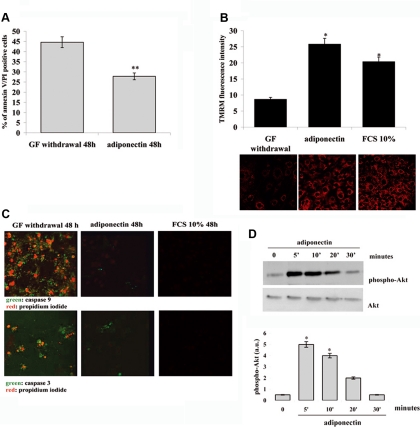
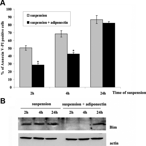
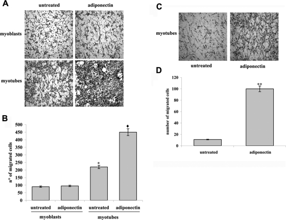
Mesoangioblasts are progenitor endowed with multipotent mesoderm differentiation ability. Despite the promising results obtained with mesoangioblast transplantation in muscle dystrophy, an improvement of their efficient engrafting and survival within damaged muscles, as well as their ex vivo activation/expansion and commitment toward myogenic lineage, is highly needed and should greatly increase their therapeutic potential. We show that globular adiponectin, an adipokine endowed with metabolic and differentiating functions for muscles, regulates vital cues of mesoangioblast cell biology. The adipokine drives mesoangioblasts to entry cell cycle and strongly counteracts the apoptotic process triggered by growth factor withdrawal, thereby serving as an activating and prosurvival stem cell factor. In addition, adiponectin provides a specific protection against anoikis, the apoptotic death due to lack of anchorage to extracellular matrix, suggesting a key protective role for these nonresident stem cells after systemic injection. Finally, adiponectin behaves as a chemoattractive factor toward mature myotubes and stimulates their differentiation toward the skeletal muscle lineage, serving as a positive regulator in mesoangioblast homing to injured or diseased muscles. We conclude that adiponectin exerts several advantageous effects on mesoangioblasts, potentially valuable to improve their efficacy in cell based therapies of diseased muscles.


Figures

Figure 1.
Figure 1.
Expression of adiponectin receptors AdipoR1 and AdipoR2 in mesoangioblasts. (A) Amount of AdipoR1 and AdipoR2 mRNA by real-time PCR. Total RNA was used for the amplification of mRNA of adiponectin receptors using as housekeeping gene 18S rRNA. The amount of target, normalized to the endogenous reference (18S RNA), was given by the 2ΔΔCT calculation and was reported as arbitrary units (a.u.). (B) Analysis of AdipoR1 and AdipoR2 expression by immunoblot. An equal amount of total proteins were run in each lane after protein assay with Bradford method, as shown by actin immunoblot. Real-time PCR is the mean of three independent assays, whereas the blot is representative of three different experiments. n.r., nonrelated band.
Figure 2.
Figure 2.
Adiponectin induces the growth of mesoangioblasts. (A) Mesoangioblasts were serum-deprived for 24 h, and where indicated, adiponectin (1 μg/ml) was added to serum-free medium for 72 h. Cells were then counted using an hemocytometer. (B) Analysis of [3H]thymidine incorporation by mesoangioblasts after the treatment with adiponectin (1 μg/ml). Cells were treated as in A, and [3H]thymidine was added during the last 2 h of incubation. These results correspond to the mean of four different experiments. *p < 0.001 and **p < 0.005 versus control. (C–E) Analysis of the signaling pathways activated by adiponectin stimulation. Mesoangioblasts were serum-deprived overnight and then stimulated with adiponectin (1 μg/ml) for the indicated period. Immunoblot analysis for the detection of the phosphorylation level of p42/p44 MAPK (Thr202/Tyr204), Akt (Ser473), p38 MAPK (Thr180/Tyr182), and AMPK (Thr182) was performed using specific phospho-antibodies. p42/p44 MAPK (C), p38 (D), and AMPK (E) immunoblots were used for normalization. Bar graph represents the phosphorylation level of the signaling proteins calculated by the ratio between the phosphorylated and total protein obtained in four different experiments. *p < 0.001 and **p < 0.005 versus time 0.
Figure 3.
Figure 3.
Adiponectin protects mesoangioblasts from apoptosis induced by GF withdrawal. Cells were serum-deprived overnight and then treated with serum-free medium with or without adiponectin (1 μg/ml) for 48 h. (A) Analysis of annexin V– and propidium iodide–positive cells by flow cytofluorimetry. (B) Analysis of mitochondria membrane potential through the staining of the cells with TMRM fluorescent probe. *p < 0.001 and **p < 0.005 versus GF withdrawal. (C) Detection of caspase-9 and -3 activation by confocal miscroscope. Mesoangioblasts were stained with FLICA probe (green) for analysis of activated caspases and with propidium iodide (red) to analyze plasma membrane modifications. (D) Phosphorylation of serine/treonine kinase Akt by adiponectin. Mesoangioblasts were serum-deprived overnight and then stimulated with adiponectin (1 μg/ml) for the indicated period. Activation of Akt was detected by immunoblot using anti-phospho-serine 473 antibodies, whereas anti-Akt antibodies were used for normalization. The Western blot is representative of three different experiments with similar results. Bar graphs in B and D are the mean of four independent experiments, whereas images are representative of three different experiments with similar results. *p < 0.001 versus time 0.
Figure 4.
Figure 4.
Adiponectin protects mesoangioblasts from anoikis. Mesoangioblasts were serum-deprived overnight and then pretreated with serum-free medium with or without adiponectin (1 μg/ml) for 12 h. Cells were then detached and maintained in suspension in serum-free medium with or without adiponectin (1 μg/ml) for 2, 4, and 24 h. (A) Apoptotic mesoangioblasts treated as described above were analyzed by annexin V and propidium iodide labeling. Bar graph is the mean of four different experiments. *p < 0.001 versus suspension. (B) Detection of Bim expression in mesoangioblasts treated as described above. The Western blot is representative of three different experiments with the same result.
Figure 5.
Figure 5.
Adiponectin enhances mesoangioblasts migration in vitro. Mesoangioblasts were serum-deprived overnight and then seeded in the upper Boyden chamber for assay. Adiponectin (1 μg/ml) was added in the upper (A and B) or lower (C and D) Boyden chamber. C2C12 myoblasts (C2C12, 0 d) or 4-d differentiated myotubes (C2C12, 4 d) were plated in the lower chamber. (A and C) Representative images of migrated mesoangioblasts after staining with hematoxylin-eosin. (B and D) Bar graph represents the mean of migrated cells counted in three different fields for each experiment. *p < 0.005 versus untreated 0 d, p < 0.001 versus adiponectin 0 d, and **p < 0.001 versus untreated.
Figure 6.
Figure 6.
Adiponectin increases skeletal muscle differentiation in vitro. Mesoangioblasts were serum-deprived overnight and then treated for 48 h with serum-free medium with or without adiponectin (1 μg/ml). The same number of mesoangioblasts was then added to a culture of rat L6 myoblasts (ratio 1:2). Myogenic differentiation was carried on for 4 d. Cells were then fixed and stained with anti-muscle myosin heavy chain (mMHC, green) and with propidium iodide (red) to label the nuclei. (A) Differentiation index of D16 mesoangioblasts per L6 coculture. (B) The graph shows the fusion index between nuclei of the murine mesoangioblasts and rat L6 myoblasts in mMHC-expressing cells with more than two nuclei compared with the total number of rat and murine nuclei. (C) Representative Western blot showing the expression of skeletal muscle markers in rat L6/murine D16 coculture after 4 d of differentiation. (D) Representative image of L6-D16 coculture after 4 d of differentiation obtained by confocal microscopy showing higher amount of myotubes in coculture containing adiponectin-treated mesoangioblasts compared with untreated. Inset shows the magnification of the myotube in the rectangle, revealing the presence of two nuclei of mesoangioblasts. The different chromatin distribution of the nuclei between rat L6 and murine D16 (arrow) is shown. *p < 0.005 versus untreated; **p < 0.001 versus untreated.
Figure 7.
Figure 7.
Adiponectin stimulates survival, chemoattraction and engrafting of mesoangioblasts in skeletal muscle of dystrophic mice. (A) Adiponectin increases mesoangioblasts survival in vivo. D16 mesoangioblasts expressing GFP were treated for 16 h with adiponectin (1 μg/ml) and then injected in the TA of Sgca-null mice. The level of expression of GFP was visualized under a stereomicroscope 3 d after injection. Bar graph shows the fluorescence intensity of GFP in lateral and medial surface of the TA muscles measured using ImageJ. Bar, 2 mm. A representative experiment is shown (n = 3). (B) Adiponectin increases the engraftment of mesoangioblasts. Top, nuclear LacZ-expressing mesoangioblasts were treated for 16 h with adiponectin and then injected in the TA of Sgca-null mice. After 24 h muscles were recovered, and a X-gal staining was performed for 4 h. Blue staining revealed an increased survival and engraftment in muscles injected with adiponectin-treated mesoangioblasts compared with control. Bar, 2 mm. A representative experiment is shown (n = 3). Bottom, 3 d after injection the muscles were recovered, and nLacZ-positive nuclei were detected by X-Gal staining. Bar, 100 μm. p = 0.028 (calculated with GraphPad). (C) Adiponectin enhances the localization of mesoangioblasts to TA of Sgca-null mice. Beads bounded with adiponectin or incubated with 0.1% BSA were injected in the TA of Sgca-null mice. The chemoattractive effect due to adiponectin was calculated as the percentage of positive GFP area near the site of injection and are reported in the plot below (arrowhead). The fluorescence intensity of GFP was measured using ImageJ software. Bar, 1 mm. A representative experiment is shown (n = 3).

Similar articles

Cited by

References

    1. Aguiari P., et al. High glucose induces adipogenic differentiation of muscle-derived stem cells. Proc. Natl. Acad. Sci. USA. 2008;105:1226–1231. - PMC - PubMed
    1. Ando Y., Inaba M., Sakaguchi Y., Tsuda M., Quan G. K., Omae M., Okazaki K., Ikehara S. Subcutaneous adipose tissue-derived stem cells facilitate colonic mucosal recovery from 2,4,6-trinitrobenzene sulfonic acid (TNBS)-induced colitis in rats. Inflamm. Bowel Dis. 2008;14:826–838. - PubMed
    1. Asakura A., Seale P., Girgis-Gabardo A., Rudnicki M. A. Myogenic specification of side population cells in skeletal muscle. J. Cell Biol. 2002;159:123–134. - PMC - PubMed
    1. Bachrach E., Li S., Perez A. L., Schienda J., Liadaki K., Volinski J., Flint A., Chamberlain J., Kunkel L. M. Systemic delivery of human microdystrophin to regenerating mouse dystrophic muscle by muscle progenitor cells. Proc. Natl. Acad. Sci. USA. 2004;101:3581–3586. - PMC - PubMed
    1. Berg A. H., Combs T. P., Scherer P. E. ACRP30/adiponectin: an adipokine regulating glucose and lipid metabolism. Trends Endocrinol. Metab. 2002;13:84–89. - PubMed

Publication types

MeSH terms

LinkOut - more resources