. 2010 Mar 26;285(13):9390-9401.
doi: 10.1074/jbc.M109.057745. Epub 2010 Jan 20.

cAMP-response element-binding protein (CREB) controls MSK1-mediated phosphorylation of histone H3 at the c-fos promoter in vitro

Affiliations

cAMP-response element-binding protein (CREB) controls MSK1-mediated phosphorylation of histone H3 at the c-fos promoter in vitro

Miho Shimada et al. J Biol Chem. .

Abstract

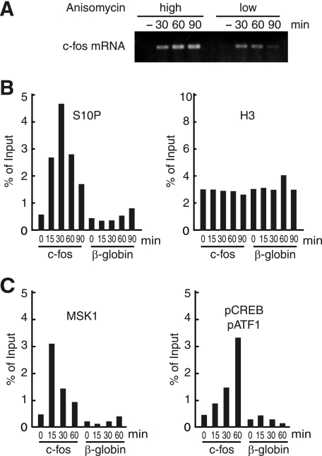
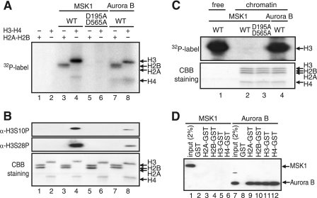
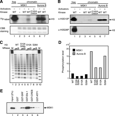
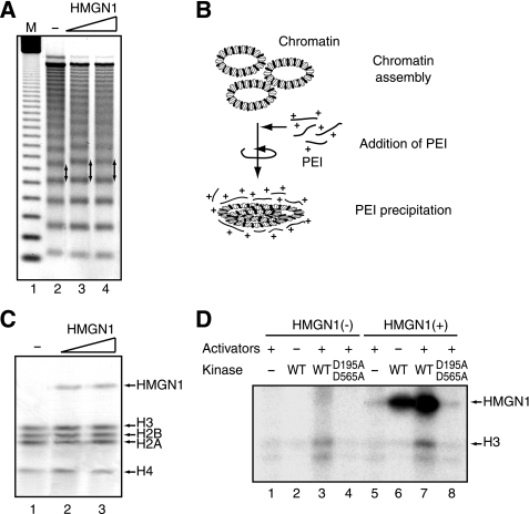
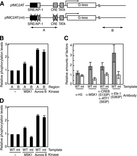
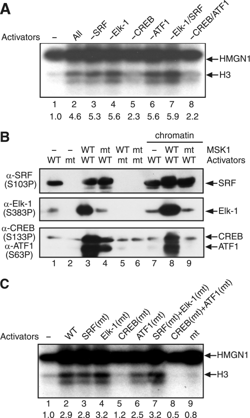
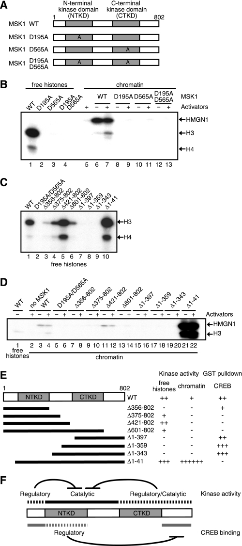
The rapid induction of the c-fos gene correlates with phosphorylations of histone H3 and HMGN1 by mitogen- and stress-activated protein kinases. We have used a cell-free system to dissect the mechanism by which MSK1 phosphorylates histone H3 within the c-fos chromatin. Here, we show that the reconstituted c-fos chromatin presents a strong barrier to histone H3 phosphorylation by MSK1; however, the activators (serum response factor, Elk-1, cAMP-response element-binding protein (CREB), and ATF1) bound on their cognate sites recruit MSK1 to phosphorylate histone H3 at Ser-10 within the chromatin. This activator-dependent phosphorylation of histone H3 is enhanced by HMGN1 and occurs preferentially near the promoter region. Among the four activators, CREB plays a predominant role in MSK1-mediated phosphorylation of histone H3, and the phosphorylation of Ser-133 in CREB is essential for this process. Mutational analyses of MSK1 show that its N-terminal inhibition domain is critical for the kinase to phosphorylate chromatin-embedded histone H3 in a CREB-dependent manner, indicating the presence of an intricate regulatory network for MSK1-mediated phosphorylation of histone H3.


Figures

FIGURE 1.
FIGURE 1.
Histone H3 phosphorylation and MSK1 recruitment on the c-fos promoter in response to anisomycin treatment. A, HeLa cells were stimulated by anisomycin with two different concentrations (high, 10 μg/ml; low, 25 ng/ml) after being starved in 0.5% fetal bovine serum for 1.5 days. The c-fos mRNA was amplified by reverse transcription-PCR, followed by electrophoresis on an agarose gel. B, HeLa cells were stimulated by anisomycin (10 μg/ml) for the indicated time and used for ChIP assays with anti-histone H3 and anti-H3-S10P antibodies. The precipitated DNA was quantified by real time PCR using primers for the c-fos gene. As control, the β-globin gene was used. The values were averaged from two independent experiments. C, occupancies of MSK1 and pCREB/pATF1 were determined by ChIP assays using antibodies against MSK1 and pCREB(Ser-133), which also recognizes pATF1(Ser-63). The values were averaged from two independent experiments.
FIGURE 2.
FIGURE 2.
MSK1 does not phosphorylate histones assembled into chromatin. A, kinase assays contained [γ-32P]ATP and 100 ng of recombinant histones with 100 ng of MSK1 (lanes 3 and 4), D195A/D565A (lanes 5 and 6), or Aurora B (lanes 7 and 8). The position of each histone is indicated on the right. WT, wild type. B, identical kinase assays were performed in the absence of [γ-32P]ATP, and the phosphorylated residues in histone H3 were identified by immunoblotting using anti-H3-S10P and anti-H3-S28P antibodies. CBB, Coomassie Brilliant Blue. C, 150 ng of chromatin assembled on pfMC2AT was phosphorylated in the presence of [γ-32P]ATP with 150 ng of MSK1 (lane 2), D195A/D565A (lane 3), or Aurora B (lane 4). As control, 10 ng of free H3-H4 tetramer were phosphorylated with MSK1 (lane 1). The positions of histone H3 on the autoradiogram (upper panel) and the core histones on the SDS-polyacrylamide gel (lower panel) are indicated on the right. D, N-terminal tails of histones fused to GST were retained on glutathione-Sepharose 4B and incubated with FLAG-tagged MSK1 (lanes 2–6) or Aurora B (lanes 8–12), and the bound proteins were analyzed by immunoblotting using anti-FLAG M2 antibody. The positions of the bound MSK1 or Aurora B are indicated on the right.
FIGURE 3.
FIGURE 3.
MSK1 phosphorylates serine 10 of histone H3 within chromatin in an activator-dependent manner. A, 150 ng of the c-fos chromatin was incubated in the presence of 150 ng of MSK1 (lanes 3–5) or Aurora B (lanes 6 and 7) and 100 ng of the four activators (lanes 4, 5, and 7) using 10 mm [γ-32P]ATP. D195A/D595A is a kinase-deficient MSK1 (lane 5). 20 ng of free H3/H4 tetramer was phosphorylated by 400 ng of MSK1 as control (lane 1). The gel was stained in Coomassie Brilliant Blue (CBB) (lower panel) and then exposed to an x-ray film (upper panel). WT, wild type. B, same sets of reactions as in A were performed in the absence of [γ-32P]ATP, and the phosphorylated residues were detected by immunoblotting using anti-H3-S10P (upper panel) or anti-H3-S28P (lower panel). C, chromatins assembled with wild-type (lanes 2 and 3) or mutant histone H3 whose serine was replaced with an alanine at both Ser-10 and Ser-28 (lanes 4 and 5), Ser-10 (lanes 6 and 7), or Ser-28 (lanes 8 and 9) were digested with two different concentrations of MNase. Molecular mass marker (M) is a 123-bp DNA ladder (lanes 1 and 10). D, kinase reactions were performed as in A, using the chromatin reconstituted with wild-type or mutant histones in the presence of MSK1 (black bars) or Aurora B (gray bars). The levels of phosphorylation were measured by ImageQuant software (GE Healthcare). E, GST (lane 2) or GST-fused activators (lanes 3–6) bound to glutathione-Sepharose 4B were incubated with FLAG-tagged MSK1, and the bound MSK1 was detected by immunoblotting using anti-FLAG M2 antibody. 2% of input FLAG-tagged MSK1 was loaded as control (lane 1).
FIGURE 4.
FIGURE 4.
HMGN1 enhances MSK1-mediated histone H3 phosphorylation within the c-fos chromatin. A, c-fos plasmid, pfMC2AT, was assembled into chromatin in the presence of 0 μg (lane 2), 2.75 μg (lane 3), and 5.5 μg (lane 4) of HMGN1, and then partially digested by MNase. The arrows indicate the increased spacing between nucleosomes by incorporated HMGN1. Lane 1, molecular mass marker (M). B, schematic depiction of chromatin precipitation by PEI. C, chromatin was assembled in the presence of 0 μg (lane 1), 2.75 μg (lane 2), and 5.5 μg (lane 3) of HMGN1 as in A, precipitated by PEI as in B, resolved by SDS-PAGE, and stained with Coomassie Brilliant Blue (CBB). The positions of precipitated proteins are indicated on the right. D, kinase assays were performed with the chromatin assembled in the presence (lanes 5–8) or absence (lanes 1–4) of 5.5 μg of HMGN1. The chromatin was incubated in the presence of [γ-32P]ATP with (lanes 1, 3–5, 7, and 9) or without (lanes 2 and 6) 100 ng each of activators (SRF, Elk-1, CREB, and ATF1). The reactions also contained wild-type (WT) MSK1 (lanes 2, 3, 6, and 7) or D195A/D565A (lanes 4 and 8). The positions of HMGN1 and H3 are indicated on the right.
FIGURE 5.
FIGURE 5.
Preferential phosphorylation of histone H3 near the c-fos promoter region. A, wild-type (pfMC2AT) and mutant (pfMC2AT(m)) c-fos templates used for in vitro ChIP assays and the regions amplified by PCR. Region A overlaps with the c-fos promoter, and region B is within the vector pUC19 and ∼1.5 kb downstream from the transcription start site. B, c-fos chromatin was phosphorylated by MSK1 in the presence of activators or by Aurora B and then analyzed by ChIP assays using anti-H3-S10P. The levels of H3-S10P were normalized by the values obtained with anti-histone H3 antibody. The values obtained without any kinase were set to 1. C, in vitro ChIP assays for the occupancies of MSK1 and phosphorylated activators (CREB, ATF1, and Elk-1). Wild-type (WT) and mutant (mt) c-fos templates were assembled into chromatin, and the kinase reactions were done in the presence of MSK1 and the activators. ChIP assays were performed with antibodies against histone H3, MSK1, pCREB(Ser-133) and pElk-1(Ser-383), using the primers for the region A. The anti-pCREB(Ser-133) antibody also recognizes pATF1(Ser-63). Gray and white bars indicate the relative amounts of histone H3, MSK1, and the activators on the wild-type and mutant templates, respectively. D, wild-type and mutant c-fos templates were assembled into chromatin, and the relative levels of H3-S10 phosphorylation in the presence of MSK1 or Aurora B were determined by ChIP assays using anti-H3-S10P and the primers for region A. The error bars represent S.E.
FIGURE 6.
FIGURE 6.
MSK1-mediated chromatin phosphorylation requires phosphorylation of Ser-133 in CREB. A, c-fos chromatin was phosphorylated by MSK1 in the presence of various combinations of activators. The reactions contained the following: no activator (lane 1), SRF, Elk-1, CREB, and ATF1 (lane 2); Elk-1, CREB, and ATF1 (lane 3); SRF, CREB, and ATF1 (lane 4); SRF, Elk-1, and ATF1 (lane 5); SRF, Elk-1, and CREB (lane 6); CREB and ATF1 (lane 7); and SRF and Elk-1 (lane 8). The positions of histone H3 and HMGN1 are indicated on the right. B, wild-type (WT) or mutant (mt) activators were analyzed by immunoblotting using the indicated phosphospecific antibodies. The control reactions (lanes 1–6) contained either wild-type or mutant MSK1 and activators in the absence of chromatin, whereas the reactions in lanes 7–9 were performed in the presence of the c-fos chromatin. C, phosphorylation-defective mutants of SRF (lane 3), Elk-1 (lane 4), CREB (lane 5), ATF1 (lane 6), SRF and Elk-1 (lane 7), and CREB and ATF1 (lane 8) were used in place of the corresponding wild-type activator(s). Control reactions contained no activator (lane 1), wild-type activators (lane 2), and mutant activators (lane 9). The positions of histone H3 and HMGN1 are indicated on the right.
FIGURE 7.
FIGURE 7.
Multiple domains within MSK1 are required for histone H3 phosphorylation. A, D195A, D565A, or D195A/D565A has an alanine in place of an aspartate at residue(s) 195, 565, or 195 and 565, respectively. WT, wild type. B, histone H3-H4 tetramers (lanes 1–4) or reconstituted chromatin (lanes 5–13) were phosphorylated by wild-type (lanes 1, 6, and 7) or mutant MSK1 (lanes 2–4 and 8–13). Mitogen- and stress-activated kinase (MSK) was omitted from lane 5. In the chromatin kinase assays, the reactions were performed in the presence (+) or absence (−) of activators (SRF, Elk-1, CREB, and ATF1). Phosphorylated HMGN1 and histones were detected by autoradiography. The positions of HMGN1 and histone H3 and H4 are indicated on the right. C, histone H3-H4 tetramers were phosphorylated by the deletion mutants of MSK1 as depicted in E. The positions of histone H3 and H4 are indicated on the right. D, chromatin kinase assays were performed with wild-type (lanes 3 and 4) or mutant MSK1 (lanes 5–22) in the presence or absence of activators. As controls, free histone H3-H4 tetramers were phosphorylated by wild-type MSK1 (lane 1). MSK1 was omitted from lane 2. E, schematic depiction of MSK1 deletion mutants and the summarized results of C and D as well as those of GST pulldown assays between the MSK1 mutants and CREB. No phosphorylation or interaction is indicated by −, and the approximate levels of phosphorylation or interaction are indicated by the increasing number of +. F, multiple intramolecular regulations control the kinase activity of MSK1 and its interactions with CREB.

Similar articles

Cited by

References

    1. Price M. A., Hill C., Treisman R. (1996) Philos. Trans. R Soc. Lond. B Biol. Sci. 351, 551–559 - PubMed
    1. Thomson S., Mahadevan L. C., Clayton A. L. (1999) Semin. Cell Dev. Biol. 10, 205–214 - PubMed
    1. Thomson S., Clayton A. L., Hazzalin C. A., Rose S., Barratt M. J., Mahadevan L. C. (1999) EMBO J. 18, 4779–4793 - PMC - PubMed
    1. Soloaga A., Thomson S., Wiggin G. R., Rampersaud N., Dyson M. H., Hazzalin C. A., Mahadevan L. C., Arthur J. S. (2003) EMBO J. 22, 2788–2797 - PMC - PubMed
    1. Chadee D. N., Hendzel M. J., Tylipski C. P., Allis C. D., Bazett-Jones D. P., Wright J. A., Davie J. R. (1999) J. Biol. Chem. 274, 24914–24920 - PubMed

Publication types

Substances