. 2010 Mar 19;285(12):8830-9.
doi: 10.1074/jbc.M109.074344. Epub 2010 Jan 20.

A role for KAI1 in promotion of cell proliferation and mammary gland hyperplasia by the gp78 ubiquitin ligase

Affiliations

A role for KAI1 in promotion of cell proliferation and mammary gland hyperplasia by the gp78 ubiquitin ligase

Bharat Joshi et al. J Biol Chem. .

Abstract

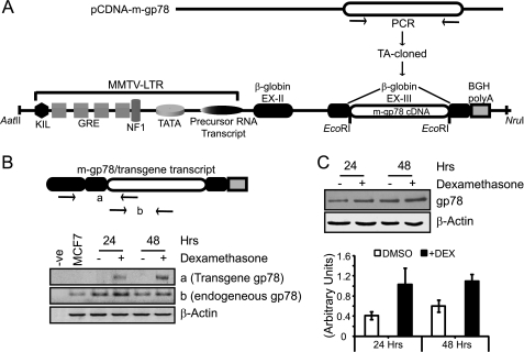
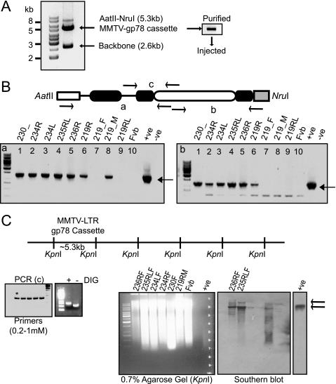
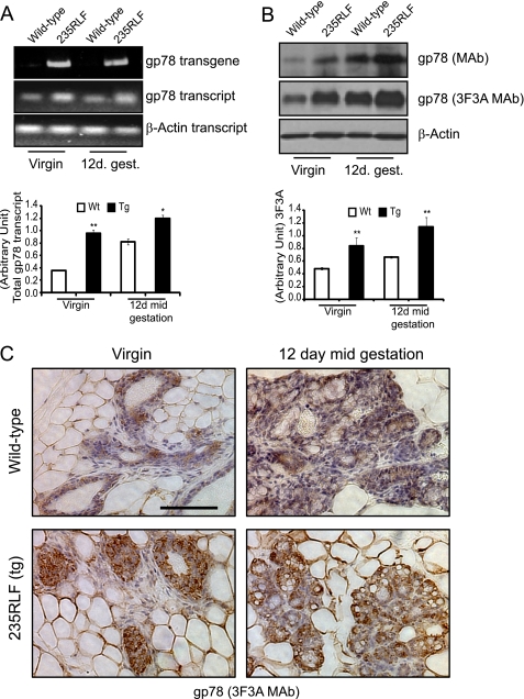
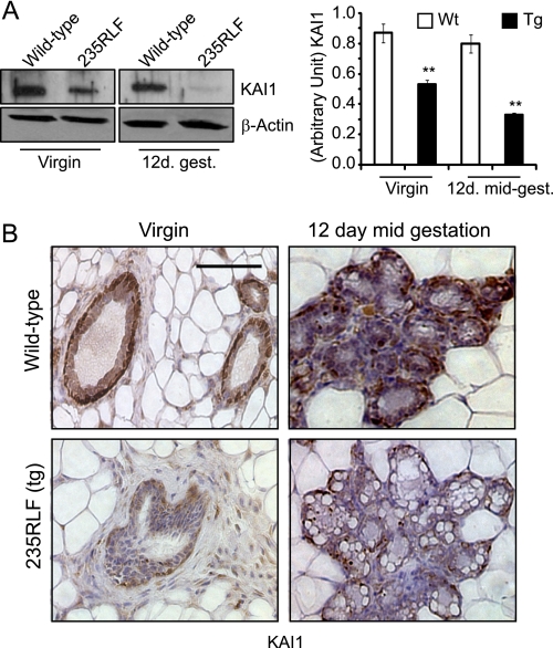
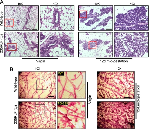
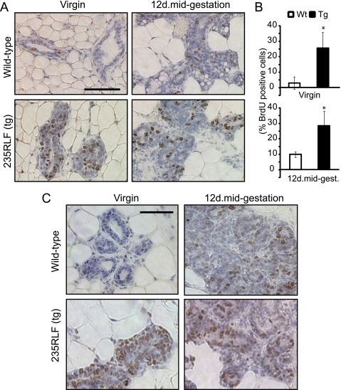
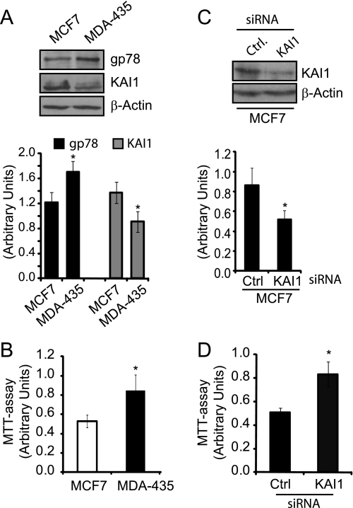
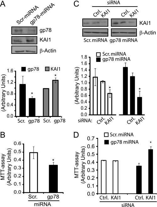
Expression of gp78, an E3 ubiquitin ligase in endoplasmic reticulum-associated degradation, is associated with tumor malignancy. To study gp78 overexpression in mammary gland development and tumorigenicity, we generated murine mammary tumor virus (MMTV) long terminal repeat-driven gp78 transgenic mice. Embryos carrying the gp78 transgene cassette were implanted in FVB surrogate mothers, and two founders with high copy integration showed elevated gp78 expression at both transcript and protein levels at the virgin stage and at 12 days gestation. Transgenic mammary glands showed increased ductal branching, dense alveolar lobule formation, and secondary terminal end bud development. Bromodeoxyuridine staining showed increased proliferation in hyperplastic ductal regions at the virgin stage and at 12 days gestation compared with wild type mice. Reduced expression of the metastasis suppressor KAI1, a gp78 endoplasmic reticulum-associated degradation substrate, demonstrates that gp78 ubiquitin ligase activity is increased in MMTV-gp78 mammary gland. Similarly, metastatic MDA-435 cells exhibit increased gp78 expression, decreased KAI1 expression, and elevated proliferation compared with nonmetastatic MCF7 cells whose proliferation was enhanced upon knockdown of KAI1. Importantly, stable gp78 knockdown HEK293 cells showed increased KAI1 expression and reduced proliferation that was rescued upon KAI1 knockdown, demonstrating that gp78 regulation of cell proliferation is mediated by KAI1. Mammary tumorigenesis was not observed in repeatedly pregnant MMTV-long terminal repeat-gp78 transgenic mice over a period of 18 months post-birth. Elevated gp78 ubiquitin ligase activity is therefore not sufficient for mammary tumorigenesis. However, the hyperplastic phenotype observed in mammary glands of MMTV-gp78 transgenic mice identifies a novel role for gp78 expression in enhancing mammary epithelial cell proliferation and nontumorigenic ductal outgrowth.


Figures

FIGURE 1.
FIGURE 1.
In vitro validation of the MMTV-LTR-gp78 plasmid. A, schematic diagram showing the design of the MMTV-LTR gp78 construct. Mouse gp78 was PCR-amplified, cloned, and placed at the EcoRI restriction site in β-globin exon III of the MMTV-LTR promoter shown with all elements. GRE represents glucocorticoid response elements. B, schematic presents gp78 transgene transcript with whole mouse gp78 fused to β-globin exons II and III and BGH-poly(A) sequence. a and b labeled on the schematic of the cassette, flanked by arrows (representing primer binding sites), show the areas used for PCR amplification. The bottom panel shows RT-PCR where gp78 transgene transcript is present only in dexamethasone-treated samples, amplified as a, and corresponds to the elevated level of total gp78 transcript, amplified as b, at both 24 and 48 h. Negative controls show no product, and control β-actin transcript levels remain unaltered. C, Western blots probed with 3F3A anti-gp78 rat IgM (1:1000) show elevation in gp78 expression upon dexamethasone treatment at both the 24- and 48-h time points. The graph represents quantification of three individual experiments showing ∼2–3-fold increase in gp78 expression levels relative to β-actin loading control (n = 3; ±S.E.; *, p < 0.05; **, p < 0.001).
FIGURE 2.
FIGURE 2.
gp78-overexpressing transgenic mice. A, the MMTV-LTR gp78 cassette (∼5.3 kb) was released from plasmid using a combination of AatII-NruI restriction enzymes, and the backbone was removed (∼2.6 kb). Cassette DNA was gel-purified, dissolved in TE at 2.5 ng/μl, and injected into superovulating 4-week-old FVB/Ntac mice. B, schematic showing released cassette with area flanked by arrows marked a, b, and c that were used for PCR genotyping. Tail genomic DNA of the obtained seven founders was assessed by PCR for transgene presence (bottom panels), and only six had complete integration of the cassette (right panel compared with left panel). Negative controls show no product, whereas a positive control using plasmid as template DNA showed amplified product of the correct size. C, schematic shows the unique restriction site KpnI used for copy number integration of the gp78 transgene cassette. Digestion produces ∼5.3-kb repeat fragments if integration of the cassette is head-to-tail. The left panel shows PCR optimization to prepare DIG-labeled PCR probe encompassing area designated as c in B, and the asterisk shows the selected condition. Labeled probe runs slower compared with nonlabeled product on agarose gel. The right panels show Southern blot genotyping where ∼10 μg of tail-genomic DNA from six founders was digested with KpnI and separated on 0.7% agarose gel along with controls (FVB genomic and MMTV-LTR-gp78 DNAs), capillary-transferred on Zeta probe (Bio-Rad) nylon membrane, and subjected to hybridization and development. The top arrow indicates genomic DNA-integrated KpnI-released cassette plus 5′ or 3′ genomic DNA extension, whereas the bottom arrow indicates an actual cassette fragment released with KpnI providing a measure of copy number of integrated cassettes. Founder lines 236RF and 235RLF show elevated levels of KpnI restriction product at the position corresponding to the positive MMTV-LTR-gp78 DNA that was absent in negative control, FVB genomic DNA, and weakly present in the rest of the founder lines.
FIGURE 3.
FIGURE 3.
gp78 overexpression in mammary gland of mouse MMTV-gp78 transgenics. A, RT-PCR analysis of mammary glands from age and estrous cycle matched wild type (Wt) and gp78 transgenic (235RLF) mice. Total RNA prepared from the pubertal virgin or 12-day mid-gestation mammary glands was subjected to cDNA synthesis and assessed for transgene and endogenous transcript levels. gp78 transgene transcript is present only in 235RLF transgenic mice (top panel). Elevated levels of total gp78 message are observed in 235RLF mammary glands compared with wild type. β-Actin transcript levels are unaltered. Quantification of total gp78 transcript normalized to β-actin is presented as a bar graph (n = 3; ±S.E.). B, 10 mg of tissue from mammary glands of wild type and gp78 transgenic mice was homogenized in 1× SDS-PAGE loading buffer, boiled for 5 min, and centrifuged, and 15 μl of lysates were loaded on 10% gel and subjected to Western blotting. gp78 expression levels probed with anti-gp78 monoclonal antibody or anti-gp78 3F3A rat IgM are increased in both the pubertal virgin and 12-day mid-gestation mammary glands of 235RLF transgenic mouse compared with age-matched wild type. Loading control β-actin shows equal loading. Quantification of three individual experiments showed 2–3-fold elevation in expression level of gp78 in the transgenic line probed with 3F3A (n = 3; ±S.E.). C, immunohistochemistry analysis of mouse mammary gland. The panels show 60× magnification of wild type as well as gp78 transgenic mouse mammary glands from pubertal virgin and 12-day mid-gestation time points stained for gp78 with 3F3A rat-IgM antibody. Increased gp78 staining of ducts and alveolar structures in transgenic glands is visible at both time points. Scale bars, 100 μm (*, p < 0.05; **, p < 0.001).
FIGURE 4.
FIGURE 4.
Reduced KAI1 expression in mammary glands of MMTV-gp78 transgenic mice. A, age-matched mammary gland lysates from wild type (Wt) or 235RLF transgenic mice from virgin or 12-day mid-gestation time points were separated on SDS-PAGE, Western blotted, and probed for KAI1. KAI1 expression levels are significantly reduced in gp78 transgenic 235RLF mice at both time points relative to wild type. Graph presents densitometric quantification (n = 3; ±S.E.). B, immunohistochemistry for KAI1 on mammary glands obtained from wild type and gp78 transgenic mouse is shown at 60× magnification. gp78 transgenic 235RLF shows a reduced level of KAI1 in ducts of transgenic mammary glands at both the virgin and 12-day mid-gestation time points compared with age-matched wild type controls. Scale bars, 100 μm (**, p < 0.001).
FIGURE 5.
FIGURE 5.
gp78 overexpression induces hyperplasia and increases ductal number and network branching in mouse mammary glands. A, hematoxylin- and eosin-stained sections (10× and 40×) show the mammary gland histology of age-matched wild type and gp78 235RLF transgenic mice. Wild type mammary glands show predominantly adipose tissue and simple epithelia in ductal or alveolar structures, whereas gp78 transgenic mammary glands show ductal alveolar hyperplasia appearing as a multi-layered epithelia protruding into the lumen, with an increased presence of vacuoles. Scale bars, 100 μm (**, p < 0.001). B, mammary ductal structure in carmine alum-stained whole mounts of age- and estrous cycle-matched wild type and transgenic mouse mammary glands shows hyperplastic, differentiated, and densely formed networks of tubules in gp78 transgenic 235RLF line compared with wild type at both the virgin and 12-day mid-gestation time points. The boxed area shows zoomed region. Similarly, 12-day mid-gestation whole mounts show significantly increased branching and alveolar development in gp78 transgenic mouse. Scale bars, 400 μm.
FIGURE 6.
FIGURE 6.
Increased cell proliferation in gp78-overexpressing mammary glands. A, mammary glands of BrdUrd-injected age-matched wild type (Wt) and gp78 235RLF transgenic mice were isolated and paraffin-embedded, and 5-μm sections were stained for BrdUrd to detect proliferating cells. B, bar graph shows quantification (means ±S.E.) of three randomly chosen fields from mammary glands of three individual transgenic or wild type mice scored for number of nuclei presenting BrdUrd incorporation relative to total nuclei. Significantly higher numbers of BrdUrd incorporated nuclei are visible in ducts and alveolar structures of 235RLF gp78 transgenic mice at the pubertal virgin and 12-day mid-gestation time points compared with wild type mice. C, ductal and alveolar epithelium of mammary glands of age-matched gp78 transgenic or wild type mice were stained with the proliferation marker Ki67. Increased Ki67 staining is present in ductal epithelia and alveolae of transgenic mammary glands compared with wild type. Scale bars, A and C, 100 μm.
FIGURE 7.
FIGURE 7.
KAI1 regulates cell proliferation in breast carcinoma cells. A, nonmetastatic MCF7 and metastatic MDA-435 breast carcinoma cell lines were Western blotted for gp78 (3F3A rat IgM), KAI1, and β-actin. Quantification of gp78 and KAI1 relative to β-actin is presented as a bar graph (n = 3; ±S.E.). B, cell proliferation of MCF7 and MDA-435 cells was determined using an MTT assay (±S.E.; n = 3; *, p < 0.05). C, MCF7 cells were transfected with KAI1-specific and control (Ctrl) siRNA and KAI1 knockdown validated by Western blot of KAI1 and β-actin. Quantification of KAI1 relative to β-actin is presented as a bar graph (n = 3; ±S.E.). D, MTT assay was performed on control and KAI1 siRNA transfected MCF7 cells (±S.E.; n = 3; *, p < 0.05).
FIGURE 8.
FIGURE 8.
KAI1 mediates gp78 regulation of cell proliferation. A, HEK293 stable cell lines expressing control (Scr, Scrambled) and gp78 targeting miRNA were Western blotted for gp78 (3F3A rat IgM), KAI1, and β-actin. Quantification of gp78 and KAI1 levels relative to β-actin is presented as a bar graph (n = 3; ±S.E.). gp78 knockdown results in increased KAI1 expression level but has no effect on β-actin expression levels. B, using the MTT assay, the stable gp78 knockdown cell line shows significantly reduced proliferation compared with control (Ctrl) cell line (±S.E.; n = 3; *, p < 0.05). C, HEK293 stable cell lines expressing control and gp78 targeting miRNA were transfected with control or KAI1-specific siRNA and probed by Western blot for KAI1 and β-actin. D, MTT assay was performed on HEK293 stable cell lines expressing control or gp78 targeting miRNA transfected with either control or KAI1-specific siRNA. KAI1 siRNA increased cell proliferation only in gp78 miRNA stable HEK293 knockdown cells (±S.E.; n = 3; *, p < 0.05).

Similar articles

Cited by

References

    1. Fairbank M., St-Pierre P., Nabi I. R. (2009) Mol. Biosyst. 5, 793–801 - PubMed
    1. Chiu C. G., St-Pierre P., Nabi I. R., Wiseman S. M. (2008) Expert. Rev. Anticancer Ther. 8, 207–217 - PubMed
    1. Sjöblom T., Jones S., Wood L. D., Parsons D. W., Lin J., Barber T. D., Mandelker D., Leary R. J., Ptak J., Silliman N., Szabo S., Buckhaults P., Farrell C., Meeh P., Markowitz S. D., Willis J., Dawson D., Willson J. K., Gazdar A. F., Hartigan J., Wu L., Liu C., Parmigiani G., Park B. H., Bachman K. E., Papadopoulos N., Vogelstein B., Kinzler K. W., Velculescu V. E. (2006) Science 314, 268–274 - PubMed
    1. Hirono Y., Fushida S., Yonemura Y., Yamamoto H., Watanabe H., Raz A. (1996) Br. J. Cancer 74, 2003–2007 - PMC - PubMed
    1. Nakamori S., Watanabe H., Kameyama M., Imaoka S., Furukawa H., Ishikawa O., Sasaki Y., Kabuto T., Raz A. (1994) Cancer 74, 1855–1862 - PubMed

Publication types

LinkOut - more resources