. 2010 Mar 26;285(13):9545-9555.
doi: 10.1074/jbc.M109.038364. Epub 2010 Jan 19.

Structure of the antibiotic resistance factor spectinomycin phosphotransferase from Legionella pneumophila

Affiliations

Structure of the antibiotic resistance factor spectinomycin phosphotransferase from Legionella pneumophila

Desiree H Fong et al. J Biol Chem. .

Abstract

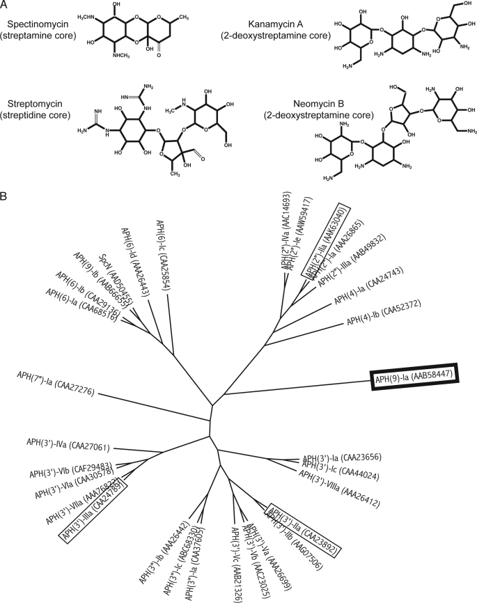
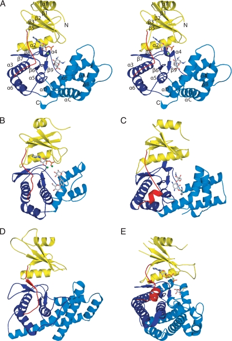
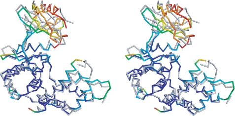
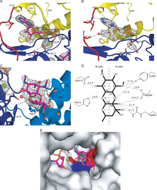
Aminoglycoside phosphotransferases (APHs) constitute a diverse group of enzymes that are often the underlying cause of aminoglycoside resistance in the clinical setting. Several APHs have been extensively characterized, including the elucidation of the three-dimensional structure of two APH(3') isozymes and an APH(2'') enzyme. Although many APHs are plasmid-encoded and are capable of inactivating numerous 2-deoxystreptmaine aminoglycosides with multiple regiospecificity, APH(9)-Ia, isolated from Legionella pneumophila, is an unusual enzyme among the APH family for its chromosomal origin and its specificity for a single non-2-deoxystreptamine aminoglycoside substrate, spectinomycin. We describe here the crystal structures of APH(9)-Ia in its apo form, its binary complex with the nucleotide, AMP, and its ternary complex bound with ADP and spectinomycin. The structures reveal that APH(9)-Ia adopts the bilobal protein kinase-fold, analogous to the APH(3') and APH(2'') enzymes. However, APH(9)-Ia differs significantly from the other two types of APH enzymes in its substrate binding area and that it undergoes a conformation change upon ligand binding. Moreover, kinetic assay experiments indicate that APH(9)-Ia has stringent substrate specificity as it is unable to phosphorylate substrates of choline kinase or methylthioribose kinase despite high structural resemblance. The crystal structures of APH(9)-Ia demonstrate and expand our understanding of the diversity of the APH family, which in turn will facilitate the development of new antibiotics and inhibitors.


Figures

FIGURE 1.
FIGURE 1.
Diversity in aminoglycoside antibiotics and the phosphotransferases that inactivate them. A, structures of representative aminoglycosides from different classes. B, cladogram for aminoglycoside phosphotransferases (APHs). Thirty-one amino acid sequences of APHs were aligned using ClustalX version 2.0 (60) and the radial style tree was produced using FigTree version 1.2.3. GenBank protein codes are noted in parentheses. APH(9)-Ia is highlighted in a box with thick black line and the other APHs whose crystal structures have been determined (APH(3′)-IIa/IIIa and APH(2″)-IIa are highlighted in boxes with thin black lines.
FIGURE 2.
FIGURE 2.
Schematic representation of APH(9)-Ia and its structural relatives. A, stereo image of the crystal structure of the ternary complex of APH(9)-Ia with ADP and spectinomycin in schematic representation. The N-terminal lobe is colored yellow, the linker between the N- and C-terminal lobe is colored red, the core region of the C-terminal lobe is colored dark blue, and the thumb is colored in light blue. The ADP and spectinomycin are shown as light gray sticks and the magnesium ion is shown in green. B, ternary complex of APH(3′)-IIIa with ADP and kanamycin (PDB code 1L8T); C, APH(2″)-IIa bound with gentamicin (PDB code 3HAM); D, apo form of CK (PDB code 2CKO); and E, ternary complex of MTRK with AMP-PNP and 5-methylthioribose (PDB code 2PUN). The coloring scheme for panels B–E is the same as panel A.
FIGURE 3.
FIGURE 3.
Stereo image of the comparison of the open and closed conformations of APH(9)-Ia. Representative open (apo, monomer A) and closed (nucleotide-bound) conformations of APH(9)-Ia are superposed using residues 108–323. The open conformation in backbone representation is colored in gray. The degree of movement in the main chain atoms as the enzyme shifts from open to closed conformation upon the binding of the nucleotide is demonstrated by a rainbow gradient of colors ranging, where blue represents no significant displacement and red represents displacements greater than 8 Å between corresponding main chain atoms. Root mean square deviation values were not calculated for sections of the structures not modeled in either the apo or AMP-bound APH(9)-Ia.
FIGURE 4.
FIGURE 4.
Active site of APH(9)-Ia. A and B, schematic representations of the nucleotide-binding site of the ternary (A) and binary (B) complexes of APH(9)-Ia. ADP is shown as magenta sticks in A and AMP is shown as white sticks in B. The protein is colored according to the assignment in Fig. 2. Interactions between amino acid residues and the nucleotide are shown in dashed lines. Lys-52 is semitransparent because its interaction with the nucleotide cannot be confirmed. Solvent molecules are shown as red spheres and the magnesium ion as a light green sphere. C, schematic representation of the substrate-binding site in the ternary complex of APH(9)-Ia. Spectinomycin is shown in magenta sticks and residues interacting with the substrate and their hydrogen bond interactions with the substrate are indicated as in A and B. D, schematic representation of the hydrogen bonding interactions between spectinomycin and APH(9)-Ia. E, molecular surface of APH(9)-Ia in the vicinity of the active site illustrating the contoured spectinomycin-binding groove. ADP and spectinomycin are shown as magenta sticks. The surface is in light gray and the polar patches surrounding spectinomycin are colored red to represent negatively charged areas and blue to represent positively charged areas. The amino acids that make direct interactions with spectinomycin are mapped to the charged patches.
FIGURE 5.
FIGURE 5.
Comparison of nucleotide binding in APH(9)-Ia, APH(3′)-IIIa, and protein kinase A. A, APH(9)-Ia, colored in magenta, and APH(3′)-IIIa, colored in green, and (B) APH(9)-Ia and protein kinase A (PDB code 1RDQ), colored in cyan, are superposed with the conserved amino acid residues to illustrate the orientation of the adenine ring in the nucleotide. The conserved residues and the nucleotides are shown in sticks and the magnesium ions are shown as spheres.

Similar articles

Cited by

References

    1. Mingeot-Leclercq M. P., Glupczynski Y., Tulkens P. M. (1999) Antimicrob. Agents Chemother. 43, 727–737 - PMC - PubMed
    1. Shaw K. J., Rather P. N., Hare R. S., Miller G. H. (1993) Microbiol. Rev. 57, 138–163 - PMC - PubMed
    1. Vakulenko S. B., Mobashery S. (2003) Clin. Microbiol. Rev. 16, 430–450 - PMC - PubMed
    1. Wright G. D., Thompson P. R. (1999) Front. Biosci. 4, D9–21 - PubMed
    1. McKay G. A., Wright G. D. (1995) J. Biol. Chem. 270, 24686–24692 - PubMed

Publication types