

Bevacizumab suppression of establishment of micrometastases in experimental ocular melanoma
- PMID: 20089875
- PMCID: PMC2874122
- DOI: 10.1167/iovs.09-4755
Bevacizumab suppression of establishment of micrometastases in experimental ocular melanoma
Abstract
Purpose: This study was undertaken to determine whether anti-vascular endothelial growth factor (VEGF) therapy inhibits growth of primary uveal melanoma and spread of its hepatic micrometastases.
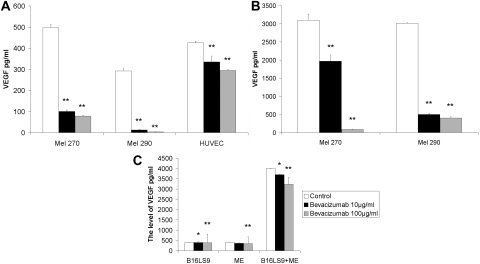
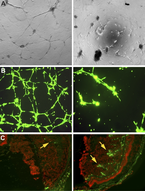
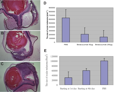
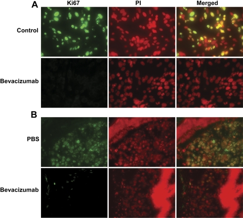
Methods: The human uveal melanoma cell lines Mel290 and Mel 270, HUVECs, mouse B16LS9 melanoma cells, and mouse vascular endothelial cells were separately cultured or co-cultured and incubated with bevacizumab or IgG1. The level of VEGF protein in the culture medium was measured by ELISA. In vitro angiogenesis and invasion assays were performed under bevacizumab or IgG1 treatment. Mel290 or B16LS9 cells were inoculated into NU/NU or C57Bl/6 mouse eyes which were enucleated after 7 days. The sizes of the intraocular tumors were determined. Time and dosage experiments were performed by using 50 or 250 microg bevacizumab starting at day 1 or 4 after inoculation. Hepatic micrometastases were enumerated. Proliferation, apoptosis, and angiogenesis markers were detected in the ocular tumor by immunofluorescence staining.
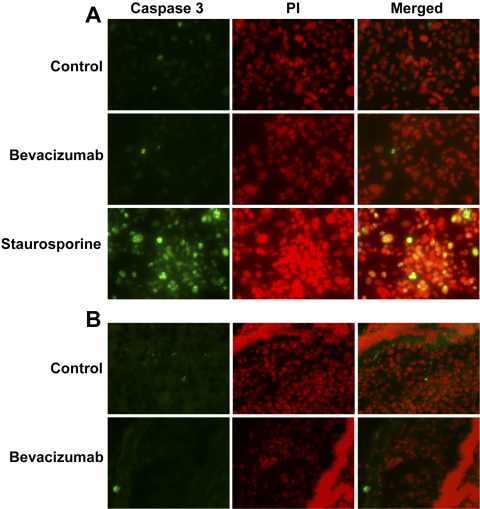
Results: Bevacizumab significantly reduced the level of VEGF in the culture media from human uveal melanoma cells, mouse melanoma cells, and co-cultured cells. It also inhibited cell tube formation and decreased in vitro invasion of tumor cells. In the mouse model, bevacizumab suppressed primary ocular melanoma growth and the formation of hepatic micrometastases in a dose-dependent manner. Furthermore, immunohistochemical staining showed decreased Ki67 and unchanged caspase 3 expression after treatment with bevacizumab.
Conclusions: Treatment with bevacizumab suppressed in vitro growth and in vivo hepatic micrometastasis of ocular melanoma cells. Bevacizumab is a potential therapeutic agent for the treatment of uveal melanoma micrometastases.
Figures
Comment in
-
Bevacizumab suppression of establishment of micrometastases in experimental ocular melanoma.Invest Ophthalmol Vis Sci. 2010 Dec;51(12):6906; author reply 6906-7. doi: 10.1167/iovs.10-6275. Invest Ophthalmol Vis Sci. 2010. PMID: 21123801 Free PMC article. No abstract available.
References
-
- Economou MA. Introduction: uveal melanoma. Acta Ophthalmol 2008;83:7–19 - PubMed
-
- Pyrhonen S. The treatment of metastatic uveal melanoma. Eur J Cancer 1998;34(suppl 3):27–30 - PubMed
-
- Yang H, Xu Z, Iuvone PM, Grossniklaus HE. Angiostatin decreases cell migration and vascular endothelium growth factor (VEGF) to pigment epithelium derived factor (PEDF) RNA ratio in vitro and in a murine ocular melanoma model. Mol Vis 2006;12:511–517 - PubMed
-
- Yang H, Akor C, Dithmar S, Grossniklaus HE. Low dose adjuvant angiostatin decreases hepatic micrometastasis in murine ocular melanoma model. Mol Vis 2004;10:987–995 - PubMed
-
- Brychtova S, Bezdekova M, Brychta T, et al. The role of vascular endothelial growth factors and their receptors in malignant melanomas. Neoplasia 2008;55:273–279 - PubMed
Publication types
MeSH terms
Substances
Grants and funding
LinkOut - more resources
Full Text Sources
Medical
Research Materials
