. 2010 Jan 20;30(3):996-1002.
doi: 10.1523/JNEUROSCI.1619-09.2010.

In vivo diffusion tensor imaging and histopathology of the fimbria-fornix in temporal lobe epilepsy

Affiliations

In vivo diffusion tensor imaging and histopathology of the fimbria-fornix in temporal lobe epilepsy

Luis Concha et al. J Neurosci. .

Abstract

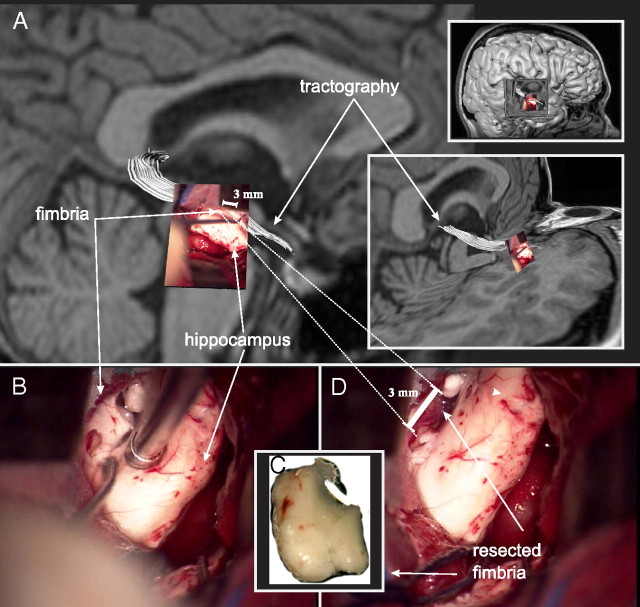
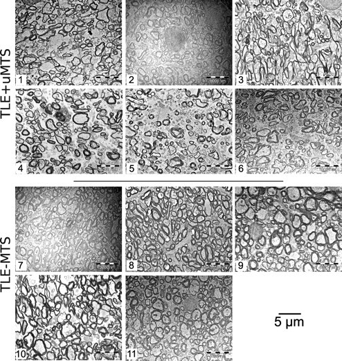
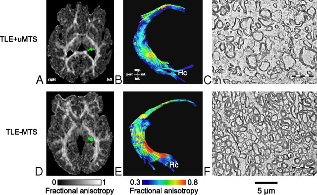
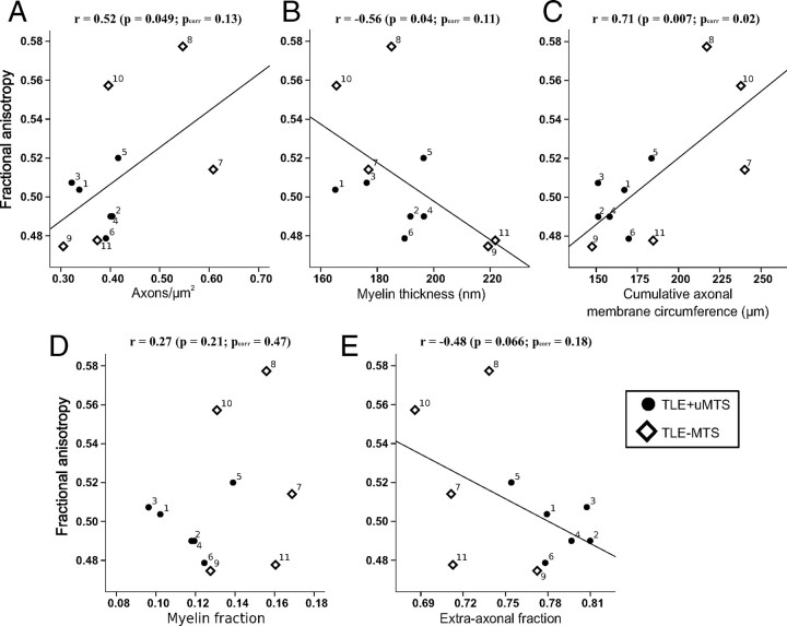
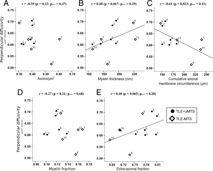
While diffusion tensor imaging (DTI) has been extensively used to infer micro-structural characteristics of cerebral white matter in human conditions, correlations between human in vivo DTI and histology have not been performed. Temporal lobe epilepsy (TLE) patients with mesial temporal sclerosis (MTS) have abnormal DTI parameters of the fimbria-fornix (relative to TLE patients without MTS) which are presumed to represent differences in axonal/myelin integrity. Medically intractable TLE patients who undergo temporal lobe resection including the fimbria-fornix provide a unique opportunity to study the anatomical correlates of water diffusion abnormalities in freshly excised tissue. Eleven patients with medically intractable TLE were recruited (six with and five without MTS) for presurgical DTI followed by surgical excision of a small specimen of the fimbria-fornix which was processed for electron microscopy. Blinded quantitative analysis of the microphotographs included axonal diameter, density and area, cumulative axon membrane circumference, and myelin thickness and area. As predicted by DTI the fimbria-fornix of TLE patients with MTS had increased extra-axonal fraction, and reduced cumulative axonal membrane circumference and myelin area. Consistent with the animal literature, water diffusion anisotropy over the crus of the fimbria-fornix was strongly correlated with axonal membranes (cumulative membrane circumference) within the surgical specimen (approximately 15% of what was analyzed with DTI). The demonstration of a correlation between histology and human in vivo DTI, in combination with the observation that in vivo DTI accurately predicted white matter abnormalities in a human disease condition, provides strong validation of the application of DTI as a noninvasive marker of white matter pathology.


Figures

Figure 1.
Figure 1.
Presurgical tractography and surgical resection of the fimbria-fornix. A photograph of the fimbria-fornix, as seen through the surgical microscope, is overlaid on the preoperative tractography of the same white matter structure (white) (A). The fimbria-fornix lies directly above the surgical instrument and the hippocampus as it is being resected (B). The specimen has been removed (C), leaving a hollow mark above the hippocampus (D). The resected specimen is immediately fixed and processed for electron microscopy. Note that the length of the crus of the fimbria-fornix analyzed with DTI (∼20 mm) is much larger than the resected specimen used for the electron microscopic quantitative histological analysis (∼3 mm, dashed lines).
Figure 2.
Figure 2.
Electron microphotographs of the fimbria-fornix. For each of the six TLE+uMTS and five TLE−MTS patients, one of the 10 available electron microscopy fields is displayed at a magnification of 3500×. Patients with mesial temporal sclerosis show fewer axons and increased extra-axonal space. Patients are identified by numbers (Table 1). Patients 5 and 8 are shown in Figure 3.
Figure 3.
Figure 3.
Electron microscopy and tractography of the fimbria-fornix. Histological fields of the fimbria-fornix resected during epilepsy surgery from two representative patients with TLE are shown with their corresponding axial FA maps (A, D, with the left fimbria-fornix marked as green) and tractography of the fimbria-fornix (B, E). The patient with mesial temporal sclerosis (Patient 5) shows lower diffusion anisotropy of the fimbria-fornix (B) than the TLE−MTS (Patient 8) (E). This corresponds to lower axonal density and higher extra-axonal fraction (C) than in the subject with TLE−MTS (F).
Figure 4.
Figure 4.
Histological correlates of fractional anisotropy in the fimbria-fornix versus axon density (A), myelin thickness (B), cumulative axonal membrane circumference (C), myelin fraction (D), and extra-axonal fraction (E). The average fractional anisotropy from the entire ipsilateral crus of the fimbria-fornix and histological parameters from electron microscopy of the smaller specimen of the fimbria-fornix adjacent to the hippocampus are plotted for each subject. FA shows the strongest correlation with cumulative axonal membrane circumference (C), namely a positive correlation indicating that anisotropy increases with greater surface area of the axonal membranes. Numbers identify each patient (Table 1).
Figure 5.
Figure 5.
Histological correlates of perpendicular diffusivity in the fimbria-fornix versus axon density (A), myelin thickness (B), cumulative membrane circumference (C), myelin fraction (D), and extra-axonal fraction (E). Cumulative axonal membrane circumference (C) from electron microscopy of the smaller specimen of the fimbria-fornix adjacent to the hippocampus shows a trend toward a negative correlation with perpendicular diffusivity from the entire ipsilateral crus of the fimbria-fornix. The perpendicular diffusion appears to be driving the anisotropy changes reported in Figure 4C. Numbers identify each patient (Table 1).

Similar articles

Cited by

References

    1. Arfanakis K, Hermann BP, Rogers BP, Carew JD, Seidenberg M, Meyerand ME. Diffusion tensor MRI in temporal lobe epilepsy. Magn Reson Imaging. 2002;20:511–519. - PubMed
    1. Basser PJ, Mattiello J, LeBihan D. MR diffusion tensor spectroscopy and imaging. Biophys J. 1994;66:259–267. - PMC - PubMed
    1. Beaulieu C. The basis of anisotropic water diffusion in the nervous system-a technical review. NMR Biomed. 2002;15:435–455. - PubMed
    1. Beaulieu C, Allen PS. Determinants of anisotropic water diffusion in nerves. Magn Reson Med. 1994a;31:394–400. - PubMed
    1. Beaulieu C, Allen PS. Water diffusion in the giant axon of the squid: implications for diffusion-weighted MRI of the nervous system. Magn Reson Med. 1994b;32:579–583. - PubMed

Publication types