. 2010 Jan 20;30(3):1096-109.
doi: 10.1523/JNEUROSCI.2309-09.2010.

Alpha2-adrenoceptor blockade accelerates the neurogenic, neurotrophic, and behavioral effects of chronic antidepressant treatment

Affiliations

Alpha2-adrenoceptor blockade accelerates the neurogenic, neurotrophic, and behavioral effects of chronic antidepressant treatment

Sudhirkumar U Yanpallewar et al. J Neurosci. .

Abstract

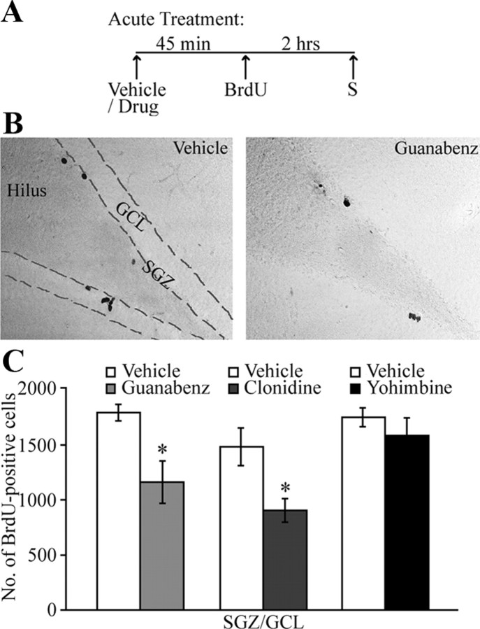
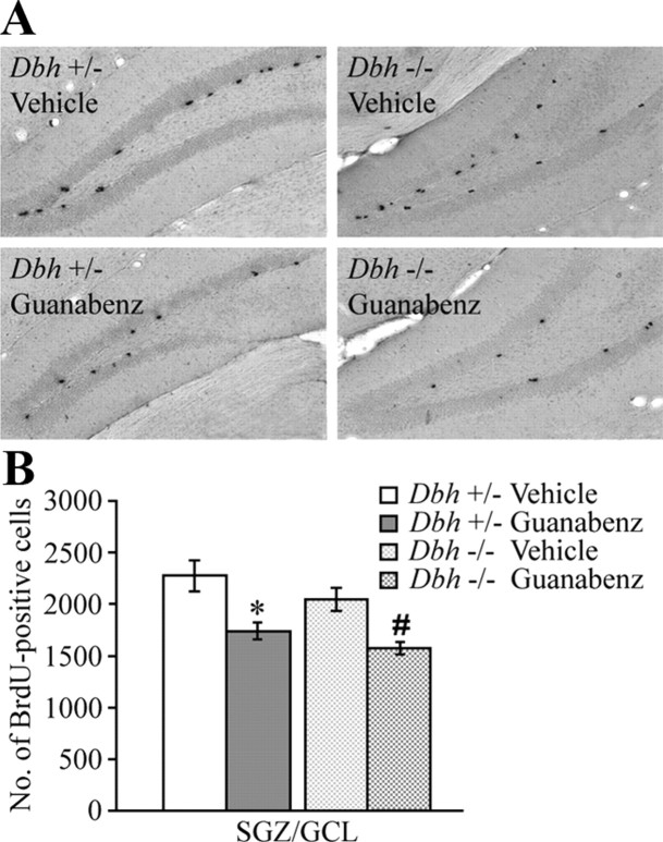
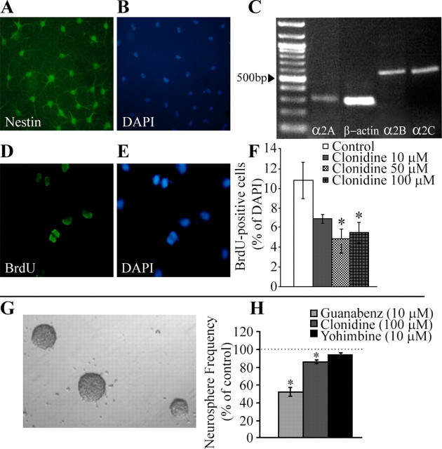
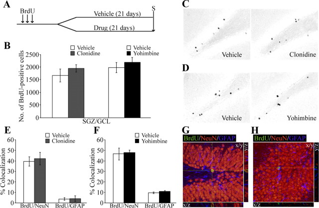
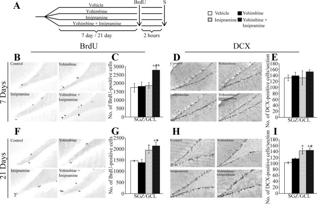
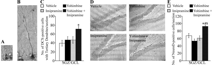
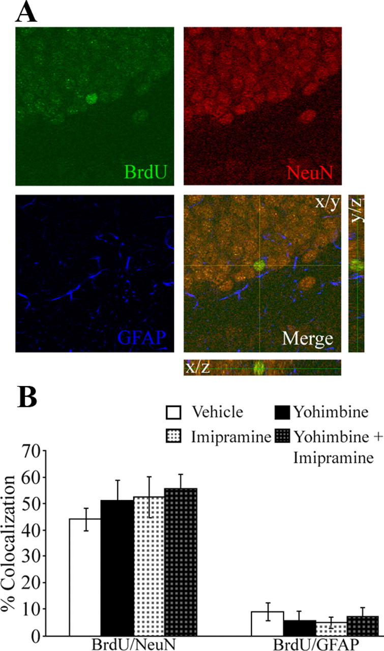
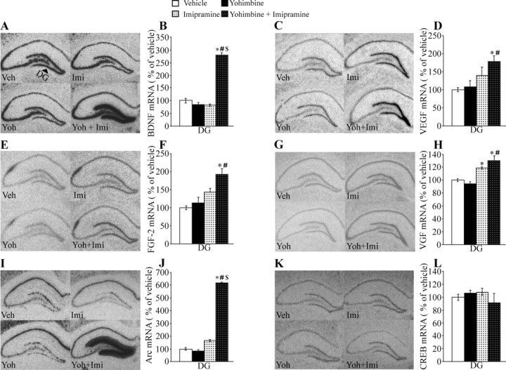
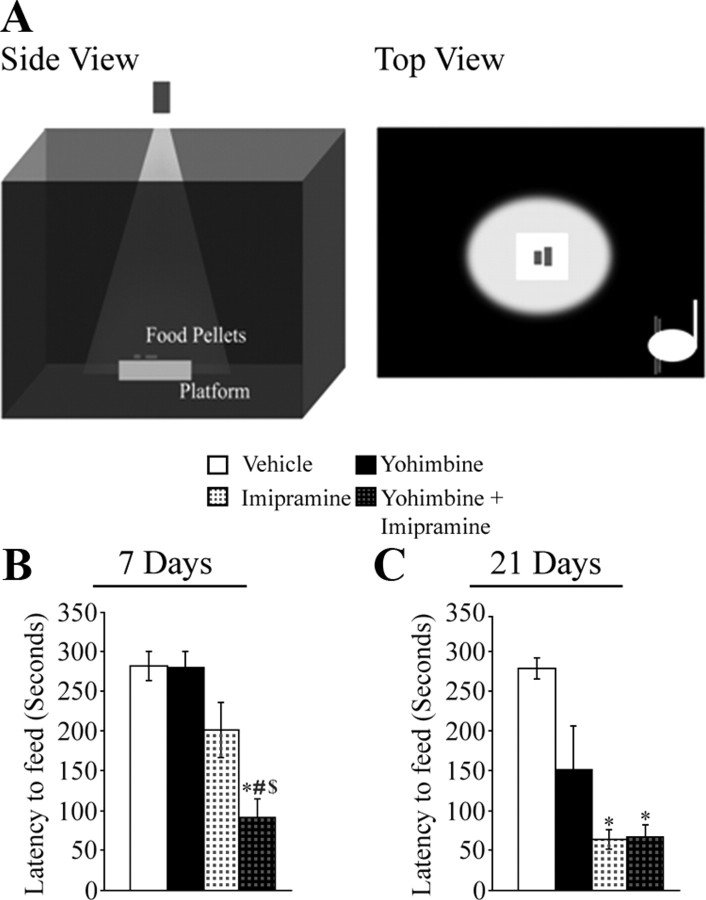
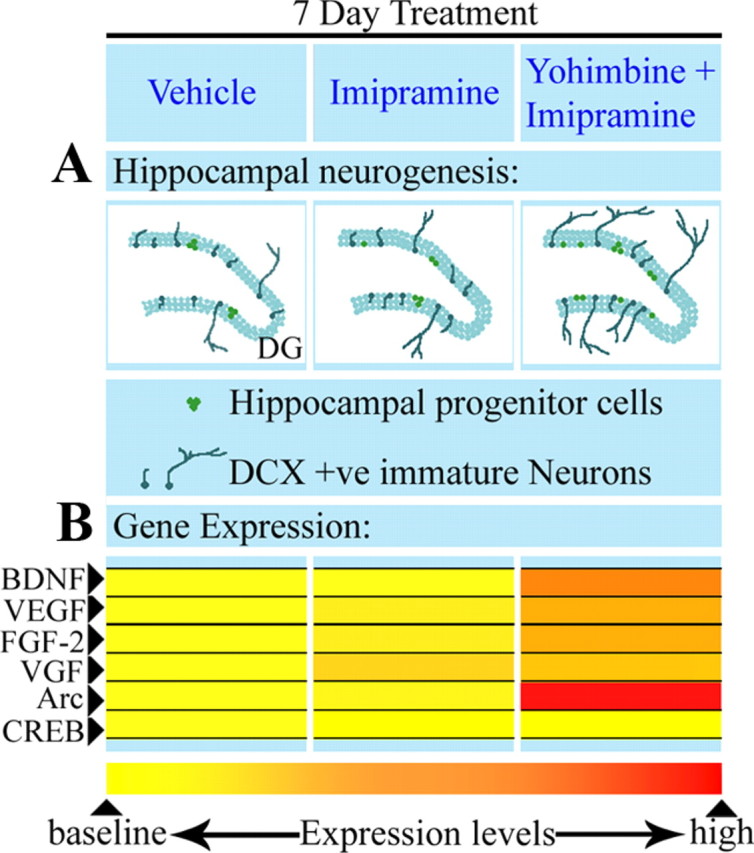
Slow-onset adaptive changes that arise from sustained antidepressant treatment, such as enhanced adult hippocampal neurogenesis and increased trophic factor expression, play a key role in the behavioral effects of antidepressants. alpha(2)-Adrenoceptors contribute to the modulation of mood and are potential targets for the development of faster acting antidepressants. We investigated the influence of alpha(2)-adrenoceptors on adult hippocampal neurogenesis. Our results indicate that alpha(2)-adrenoceptor agonists, clonidine and guanabenz, decrease adult hippocampal neurogenesis through a selective effect on the proliferation, but not the survival or differentiation, of progenitors. These effects persist in dopamine beta-hydroxylase knock-out (Dbh(-/-)) mice lacking norepinephrine, supporting a role for alpha(2)-heteroceptors on progenitor cells, rather than alpha(2)-autoreceptors on noradrenergic neurons that inhibit norepinephrine release. Adult hippocampal progenitors in vitro express all the alpha(2)-adrenoceptor subtypes, and decreased neurosphere frequency and BrdU incorporation indicate direct effects of alpha(2)-adrenoceptor stimulation on progenitors. Furthermore, coadministration of the alpha(2)-adrenoceptor antagonist yohimbine with the antidepressant imipramine significantly accelerates effects on hippocampal progenitor proliferation, the morphological maturation of newborn neurons, and the increase in expression of brain derived neurotrophic factor and vascular endothelial growth factor implicated in the neurogenic and behavioral effects of antidepressants. Finally, short-duration (7 d) yohimbine and imipramine treatment results in robust behavioral responses in the novelty suppressed feeding test, which normally requires 3 weeks of treatment with classical antidepressants. Our results demonstrate that alpha(2)-adrenoceptors, expressed by progenitor cells, decrease adult hippocampal neurogenesis, while their blockade speeds up antidepressant action, highlighting their importance as targets for faster acting antidepressants.


Figures

Figure 1.
Figure 1.
α2-Adrenoceptor stimulation decreases the proliferation of adult hippocampal progenitors. Rats received acute treatment with the α2-adrenoceptor agonists guanabenz or clonidine, or the α2-adrenoceptor antagonist yohimbine as described in Materials and Methods. A, Shown is a schematic representation of the experimental design to assess the influence of acute treatment with α2-adrenoceptor agonists and antagonist on adult hippocampal progenitor proliferation (S-time point for kill). C, Quantitative stereological analysis revealed a significant decrease in the number of BrdU-positive cells in the SGZ/GCL following acute treatment with the α2-adrenoceptor agonists guanabenz and clonidine. B, Shown are representative photomicrographs of BrdU-positive cells from vehicle- and acute guanabenz-treated groups. BrdU-positive cells were observed in the SGZ at the border of the GCL and the hilus. Acute treatment with the α2-adrenoceptor antagonist yohimbine did not alter the number of BrdU-positive cells in the SGZ/GCL (C). The results are expressed as the mean ± SEM number of BrdU-positive cells in the SGZ/GCL (n = 5–6 per group). *p < 0.05 compared with vehicle-treated controls (Student's t test).
Figure 2.
Figure 2.
α2-Adrenoceptor stimulation-induced decline in adult hippocampal progenitor proliferation persists in Dbh−/− mice. Dbh+/− and Dbh−/− mice received treatment with guanabenz or vehicle once daily for 7 d as described in Materials and Methods. A, Shown are representative photomicrographs of BrdU-positive cells from vehicle- and guanabenz-treated Dbh+/− and Dbh−/− animals. B, Quantitative analysis indicated a significant reduction in BrdU-positive cell number within the SGZ/GCL of both Dbh+/− and Dbh−/− mice following guanabenz treatment. The results are expressed as the mean ± SEM number of BrdU-positive cells in the SGZ/GCL (n = 5–6 per group). *p < 0.05 compared with Dbh+/− vehicle; #p < 0.05 compared with Dbh−/− vehicle (ANOVA and Bonferroni post hoc test).
Figure 3.
Figure 3.
α2-Adrenoceptor stimulation decreases the proliferation of adult hippocampal progenitors in vitro in two culture models, dispersed adult hippocampal progenitor cultures and the Neurosphere assay. A, B, Nestin-immunopositive (A, Nestin; B, DAPI), dispersed adult hippocampal progenitors were derived as described in Materials and Methods and in the study by Palmer et al. (1999). C, Reverse transcriptase-PCR results indicated that adult hippocampal progenitors express mRNA for the α2A-, α2B-, and α2C-adrenoceptor subtypes. F, Treatment of dispersed hippocampal progenitors with clonidine (10, 50, or 100 μm) resulted in a significant decline in BrdU incorporation at the 50 and 100 μm doses. D, E, Shown is an epifluorescent image of BrdU-immunopositive progenitor cells (D, BrdU; E, DAPI). α2-Adrenoceptor stimulation also inhibited the proliferation of neural stem cells in vitro as observed using the neurosphere assay. The neurosphere assay was performed as described in Materials and Methods. H, Treatment with the α2-adrenoceptor agonists guanabenz (10 μm) or clonidine (100 μm) resulted in decreased neurosphere formation. Treatment with the α2-adrenoceptor antagonist yohimbine (10 μm) did not alter neurosphere frequency. G, Shown is a representative image of neurospheres. Results are expressed as mean ± SEM. BrdU-positive cells as a percentage of total DAPI-positive cells (F) or mean ± SEM. Neurosphere frequency is represented as a percentage of control (H) (n = 3/group). *p < 0.05 compared with control (ANOVA and Bonferroni post hoc test).
Figure 4.
Figure 4.
α2-Adrenoceptor stimulation or blockade does not influence the survival and differentiation of adult hippocampal progenitors. Drug-naive rats first received BrdU injections followed by treatment with vehicle, clonidine or yohimbine for 21 d as described in Materials and Methods. A, Shown is a schematic representation of the experimental design to assess the influence of α2-adrenoceptor stimulation or blockade on adult hippocampal progenitor survival and differentiation (S-time point for kill). B, Quantitative analysis revealed no difference in the number of BrdU-positive cells in the SGZ/GCL following treatment with the α2-adrenoceptor agonist, clonidine or the antagonist, yohimbine. C, D, Shown are representative photomicrographs of BrdU-positive cells from clonidine- (C) and yohimbine- (D) treated groups along with representative images from respective vehicle-treated animals. The results are expressed as the mean ± SEM number of BrdU-positive cells in the SGZ/GCL (n = 5–6 per group). E, F, Quantitative analysis showed that clonidine (E) or yohimbine (F) treatment did not affect the percentage colocalization of BrdU-positive cells with either the neuronal marker NeuN or the glial marker GFAP, in the SGZ and GCL, compared with vehicle-treated controls. G, H, Shown are representative confocal z-stack images for colocalization of a BrdU-positive cell with NeuN (G) or GFAP (H). Results are expressed as the mean ± SEM percentage colocalization of BrdU-positive cells with NeuN or GFAP in the SGZ/GCL (n = 4/group).
Figure 5.
Figure 5.
α2-Adrenoceptor blockade results in faster effects of the antidepressant imipramine on adult hippocampal progenitor proliferation. Rats received combined treatment with the α2-adrenoceptor antagonist, yohimbine, and the antidepressant imipramine for 7 or 21 d as described in Materials and Methods. A, Shown is a schematic representation of the experimental design (S-time point for kill). Influence of 7 or 21 d combined yohimbine and imipramine treatments on adult hippocampal neurogenesis was assessed using the mitotic marker BrdU to determine effects on proliferation, and DCX to determine effects on immature neuron number. C, Quantitative analysis revealed a significant increase in BrdU-positive cell number in the SGZ/GCL of animals that received short-duration 7 d combined treatments with yohimbine and imipramine. Imipramine treatment by itself for 7 d did not alter BrdU-positive cell number in the SGZ/GCL (C). G, Twenty-one day treatment with imipramine, or with combined yohimbine and imipramine, resulted in a significant increase in BrdU-positive cell number in the SGZ/GCL. B, F, Shown are representative photomicrographs of BrdU-positive cells from control-, yohimbine-, imipramine-, and yohimbine + imipramine-treated groups from the 7 d (B) and 21 d (F) experiments. E, I, While in the 7 d experiment the number of DCX-positive cells per section was not altered in any of the groups (E), in the 21 d experiment imipramine, as well as combined yohimbine and imipramine, treated groups showed a significant increase in DCX-positive cells per section as compared to control (I). D, H, Shown are representative photomicrographs of DCX-positive cells from control-, yohimbine-, imipramine-, and yohimbine + imipramine-treated groups from the 7 d (D) and 21 d (H) experiments. The results are expressed as the mean ± SEM number of BrdU-positive cells per SGZ/GCL, or the mean ± SEM. Number of DCX-positive cells per section (n = 10/group). *p < 0.05 compared with control, #p < 0.05 compared with yohimbine, $p < 0.05 compared with imipramine (ANOVA and Bonferroni post hoc test).
Figure 6.
Figure 6.
α2-Adrenoceptor blockade accelerates the effect of the antidepressant imipramine on the morphological maturation of DCX-positive cells and the number of NeuroD-positive progenitors. Rats received combined treatment with the α2-adrenoceptor antagonist, yohimbine and the antidepressant, imipramine for 7 d as described in Materials and Methods. The effect of combination treatment on the morphological maturation of newborn neurons was assessed by counting the number of DCX immunopositive cells bearing a complex dendritic morphology. A, B, Shown are representative photomicrographs of a DCX-positive cell without tertiary dendrites (A) and a cell with tertiary dendrites (B). C, Quantitative analysis revealed a significant increase in the number of DCX-positive cells with tertiary dendrites/section in the SGZ/GCL of yohimbine + imipramine-treated animals compared with vehicle-treated controls. The results are expressed as the mean ± SEM number of DCX-positive cells with tertiary dendrites per section (n = 4–5/group). D, Influence of 7 d combined yohimbine and imipramine treatment on the number of NeuroD-positive progenitors was assessed using NeuroD immunohistochemistry. Shown are representative photomicrographs of NeuroD-positive cells within the SGZ/GCL of vehicle-, yohimbine-, imipramine-, and yohimbine + imipramine-treated groups. E, Quantitative analysis revealed a significant increase in the number of NeuroD-positive cells/section in the combined yohimbine and imipramine group, with no change observed following individual treatments with yohimbine or imipramine. The results are expressed as the mean ± SEM. Number of NeuroD-positive cells per section (n = 5/group). *p < 0.05 compared with control, #p < 0.05 compared with yohimbine, $p < 0.05 compared with imipramine (ANOVA and Bonferroni post hoc test).
Figure 7.
Figure 7.
Combined α2-adrenoceptor antagonist, yohimbine and antidepressant, imipramine treatment for 7 d results in an increase in adult hippocampal neurogenesis. Rats received BrdU at the end of the 7 d of combined treatment with yohimbine and imipramine, and were killed 28 d later as described in Materials and Methods. B, Confocal z-stack analysis confirmed that BrdU-positive cells differentiate primarily into NeuN-immunopositive neurons. Quantitative analysis indicated no difference in the percentage colocalization of BrdU with NeuN or GFAP across the treatment groups. A, Shown are confocal z-stack images of a BrdU-positive cell colocalizing with NeuN. The merged image demonstrates the colocalization of BrdU with NeuN. The results are expressed as the mean ± SEM percentage colocalization of BrdU-positive cells with NeuN or GFAP in the SGZ/GCL (n = 4/group).
Figure 8.
Figure 8.
Short-duration combined yohimbine and imipramine treatment for 7 d upregulates the mRNA expression of plasticity associated genes like BDNF, VEGF, FGF-2, VGF, and Arc in the dentate gyrus. Rats received treatment with the α2-adrenoceptor antagonist, yohimbine and the antidepressant, imipramine for 7 d before kill and processing for in situ hybridization as described in Materials and Methods. In situ hybridization for BDNF, VEGF, FGF-2, VGF, Arc, and CREB transcripts was performed using specific riboprobes and transcript levels in the hippocampal DG subfield were quantified using densitometric analysis. A, C, E, G, I, K, Representative autoradiograms of hippocampal sections from vehicle-, yohimbine-, imipramine-, and yohimbine + imipramine-treated animals are shown for BDNF (A), VEGF (C), FGF-2 (E), VGF (G), Arc (I) and CREB (K) mRNA. Arrowhead indicates the DG (A). Levels of mRNA in the DG hippocampal subfield are shown in the bar graphs next to the representative autoradiograms. B, D, F, H, J, Levels of BDNF (B), VEGF (D), FGF-2 (F), VGF (H) and Arc (J) mRNA in the DG were significantly increased following 7 d combined yohimbine + imipramine treatment compared with vehicle-treated controls. K, L, CREB mRNA levels were unchanged following combination treatment with yohimbine and imipramine. Results are expressed as percentage of vehicle and are the mean ± SEM (n = 4/group). *p < 0.05 compared with vehicle, #p < 0.05 compared with yohimbine, $p < 0.05 compared with imipramine (ANOVA and Bonferroni post hoc test).
Figure 9.
Figure 9.
α2-Adrenoceptor blockade hastens the effects of the antidepressant imipramine on behavior in the NSF test. Rats received combined treatment with the α2-adrenoceptor antagonist, yohimbine and the antidepressant imipramine for 7 or 21 d, and behavior was assessed using the NSF test as described in Materials and Methods. Antidepressants require chronic (2–3 weeks) administration to exhibit behavioral effects on the NSF test. A, Shown is a schematic illustration of the side, and top, views of an NSF arena in which food pellets are placed in the center on an illuminated platform and latency to feed is measured. C, Administration of imipramine or combined yohimbine + imipramine treatment for 21 d resulted a significant reduction in the latency to feed in the NSF test. Twenty-one day yohimbine treatment did not result in a significant effect on the NSF test. B, A reduced latency to feed was observed in the short-duration (7 d) experiment only in the combined yohimbine + imipramine treatment group. The results are expressed as the mean ± SEM latency to feed in seconds (n = 10/group). *p < 0.05 compared with vehicle, #p < 0.05 compared with yohimbine, $p < 0.05 compared with imipramine (ANOVA and Bonferroni post hoc test).
Figure 10.
Figure 10.
Short-duration (7 d) combined treatment with the α2-adrenoceptor antagonist, yohimbine and the antidepressant, imipramine treatment hastens the effects of antidepressant treatment on adult hippocampal progenitor proliferation, morphological maturation of newborn neurons and the expression of plasticity-associated genes. A, Shown in the illustration are the effects of combined yohimbine and imipramine treatment on adult hippocampal neurogenesis. The short-duration combined treatment results in an increased proliferation of adult hippocampal progenitors, whereas treatment with the antidepressant imipramine alone does not alter hippocampal progenitor proliferation. While the combination treatment for 7 d does not increase DCX-positive cell number, it significantly shifts the ratio of DCX-positive cells toward those bearing a more complex dendritic morphology indicating an effect of yohimbine + imipramine treatment on the morphological maturation of immature neurons. B, Shown in the illustration are the effects of combined yohimbine and imipramine treatment on the mRNA expression of trophic factors like BDNF, VEGF, and FGF-2 and plasticity-associated transcripts like Arc, VGF, and CREB. The combination treatment significantly enhances the expression of BDNF, VEGF, FGF-2, VGF and Arc in the DG subfield of the hippocampus. Effects on mRNA expression levels are schematized based on the color scale (not to exact scale).

Similar articles

Cited by

References

    1. Airan RD, Meltzer LA, Roy M, Gong Y, Chen H, Deisseroth K. High-speed imaging reveals neurophysiological links to behavior in an animal model of depression. Science. 2007;317:819–823. - PubMed
    1. Altman JD, Trendelenburg AU, MacMillan L, Bernstein D, Limbird L, Starke K, Kobilka BK, Hein L. Abnormal regulation of the sympathetic nervous system in alpha2A-adrenergic receptor knockout mice. Mol Pharmacol. 1999;56:154–161. - PubMed
    1. Andrade C, Sudha S. Electroconvulsive therapy and the alpha-2 noradrenergic receptor: implications of treatment schedule effects. J ECT. 2000;16:268–278. - PubMed
    1. Bachis A, Mallei A, Cruz MI, Wellstein A, Mocchetti I. Chronic antidepressant treatments increase basic fibroblast growth factor and fibroblast growth factor-binding protein in neurons. Neuropharmacology. 2008;55:1114–1120. - PMC - PubMed
    1. Blier P. The pharmacology of putative early-onset antidepressant strategies. Eur Neuropsychopharmacol. 2003;13:57–66. - PubMed

Publication types

MeSH terms

Substances