. 2010 Jan 20;30(3):1176-84.
doi: 10.1523/JNEUROSCI.2970-09.2010.

Rapid, learning-induced inhibitory synaptogenesis in murine barrel field

Affiliations

Rapid, learning-induced inhibitory synaptogenesis in murine barrel field

Malgorzata Jasinska et al. J Neurosci. .

Abstract

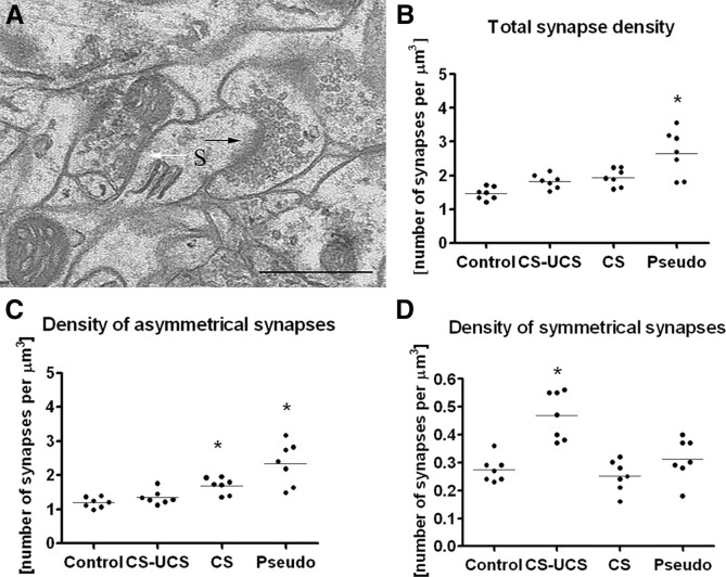
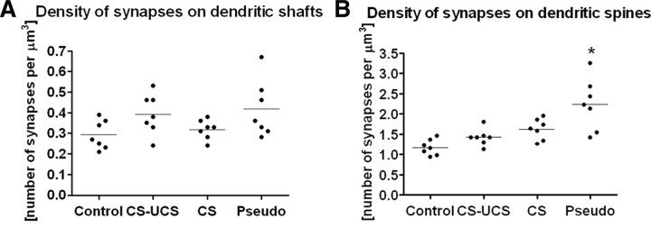
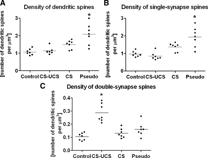
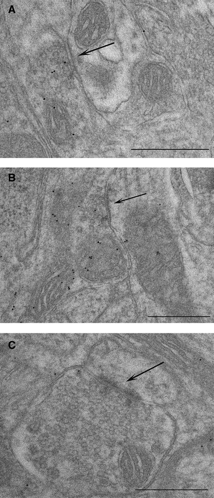
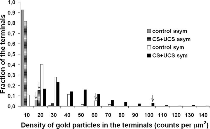
The structure of neurons changes during development and in response to injury or alteration in sensory experience. Changes occur in the number, shape, and dimensions of dendritic spines together with their synapses. However, precise data on these changes in response to learning are sparse. Here, we show using quantitative transmission electron microscopy that a simple form of learning involving mystacial vibrissae results in approximately 70% increase in the density of inhibitory synapses on spines of neurons located in layer IV barrels that represent the stimulated vibrissae. The spines contain one asymmetrical (excitatory) and one symmetrical (inhibitory) synapse (double-synapse spines), and their density increases threefold as a result of learning with no apparent change in the density of asymmetrical synapses. This effect seems to be specific for learning because pseudoconditioning (in which the conditioned and unconditioned stimuli are delivered at random) does not lead to the enhancement of symmetrical synapses but instead results in an upregulation of asymmetrical synapses on spines. Symmetrical synapses of cells located in barrels receiving the conditioned stimulus also show a greater concentration of GABA in their presynaptic terminals. These results indicate that the immediate effect of classical conditioning in the "conditioned" barrels is rapid, pronounced, and inhibitory.


Figures

Figure 1.
Figure 1.
Whisker conditioning induces a large increase in the density of symmetrical synapses (ANOVA, p < 0.001), whereas pseudoconditioning and whisker stimulation alone increased the density of asymmetrical synapses (ANOVA, p < 0.001 and p < 0.05, respectively). A, An electron micrograph taken from a B2 barrel hollow showing an asymmetrical synapse (black arrow) and a symmetrical synapse (white arrow), both located on the same dendritic spine (S). B, Total density of synapses for individual animals belonging to the following groups: control; CS–UCS, conditioned; CS, stimulated only; Pseudo, pseudoconditioned. C, Density of asymmetrical synapses for individual animals belonging to groups of treatment as in B. D, Density of symmetrical synapses for individual animals belonging to groups of treatment as in B. Scale bar, 0.5 μm. Horizontal line through the points in B–D indicates the mean.
Figure 2.
Figure 2.
Only pseudoconditioning leads to the upregulation of synapse density and only on dendritic spines (mean ANOVA, p < 0.001). A, The density of synapses on dendritic shafts does not change despite treatment. B, Pseudoconditioning increases the number of synapses on dendritic spines. Labels as in Figure 1. Horizontal line through the points indicates the mean. Each dot represents data taken from one animal.
Figure 3.
Figure 3.
Pseudoconditioning increases the density of single-synapse spines (ANOVA, p < 0.001), whereas whisker conditioning increases the density of double-synapse spines (ANOVA, p < 0.001). A, Total density of dendritic spines across treatment groups. B, Density of single-synapse spines across treatment groups. C, Density of double-synapse spines across treatment groups. Labels as in Figure 1. Horizontal line through the points indicates the mean. Each dot represents data taken from one animal.
Figure 4.
Figure 4.
Immunogold labeling for GABA. Whisker conditioning upregulates GABA in symmetrical synapse terminals as indicated by an increase in the number of gold particles per terminal. Electron micrographs taken from B2 barrel hollow stained with anti-GABA antibodies showing the following: A, symmetrical synapse from the control animal; B, symmetrical synapse from the conditioned animal; C, asymmetrical synapse from the conditioned animal. Black arrows indicate the synapses. Scale bars, 0.5 μm.
Figure 5.
Figure 5.
Quantification of immunogold labeling for GABA. Whisker conditioning upregulates GABA in the symmetrical synapse terminals. The histogram illustrates the distribution of gold particle densities in the symmetrical and asymmetrical synapse terminals (uncorrected for the background) of trained and untrained animals after pooling the data from all experimental animals. The arrowheads point to the 95th percentile of maximal density of all four density distributions to show that the substantial shift in the density of gold particles attributable to conditioning takes place only in the terminals of the symmetrical synapses (Kolmogorov–Smirnov, p < 0.001).
Figure 6.
Figure 6.
The number of head turnings decreases during conditioning and whisker stimulation alone (Mann–Whitney U test, p < 0.05) but not during pseudoconditioning. Note that, in the case of conditioned animals, changes happen already during the first session. Con, Conditioned; CS, whisker-stimulated only; Pseu, pseudoconditioned. The numbers are session numbers.

Similar articles

Cited by

References

    1. Armstrong-James M, Fox K. Spatiotemporal convergence and divergence in the rat S1 “barrel” cortex. J Comp Neurol. 1987;263:265–281. - PubMed
    1. Aroniadou-Anderjaska V, Keller A. Intrinsic inhibitory pathways in mouse barrel cortex. Neuroreport. 1996;7:2363–2368. - PubMed
    1. Bailey CH, Kandel ER. Synaptic remodeling, synaptic growth and the storage of long-term memory in Aplysia . Prog Brain Res. 2008;169:179–198. - PubMed
    1. Berry J, Krause WC, Davis RL. Olfactory memory traces in Drosophila . Prog Brain Res. 2008;169:293–304. - PMC - PubMed
    1. Costa-Mattioli M, Sonenberg N. Translational control of gene expression: a molecular switch for memory storage. Prog Brain Res. 2008;169:81–95. - PubMed

Publication types

Substances

LinkOut - more resources