

A prospective diffusion tensor imaging study in mild traumatic brain injury
- PMID: 20089939
- PMCID: PMC2830922
- DOI: 10.1212/WNL.0b013e3181d0ccdd
A prospective diffusion tensor imaging study in mild traumatic brain injury
Abstract
Objectives: Only a handful of studies have investigated the nature, functional significance, and course of white matter abnormalities associated with mild traumatic brain injury (mTBI) during the semi-acute stage of injury. The present study used diffusion tensor imaging (DTI) to investigate white matter integrity and compared the accuracy of traditional anatomic scans, neuropsychological testing, and DTI for objectively classifying mTBI patients from controls.
Methods: Twenty-two patients with semi-acute mTBI (mean = 12 days postinjury), 21 matched healthy controls, and a larger sample (n = 32) of healthy controls were studied with an extensive imaging and clinical battery. A subset of participants was examined longitudinally 3-5 months after their initial visit.
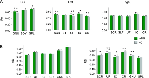
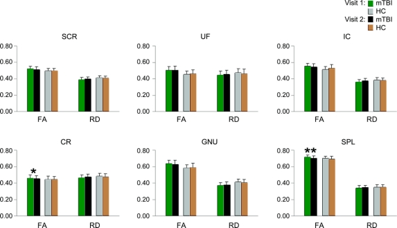
Results: mTBI patients did not differ from controls on clinical imaging scans or neuropsychological performance, although effect sizes were consistent with literature values. In contrast, mTBI patients demonstrated significantly greater fractional anisotropy as a result of reduced radial diffusivity in the corpus callosum and several left hemisphere tracts. DTI measures were more accurate than traditional clinical measures in classifying patients from controls. Longitudinal data provided preliminary evidence of partial normalization of DTI values in several white matter tracts.
Conclusions: Current findings of white matter abnormalities suggest that cytotoxic edema may be present during the semi-acute phase of mild traumatic brain injury (mTBI). Initial mechanical damage to axons disrupts ionic homeostasis and the ratio of intracellular and extracellular water, primarily affecting diffusion perpendicular to axons. Diffusion tensor imaging measurement may have utility for objectively classifying mTBI, and may serve as a potential biomarker of recovery.
Figures
Comment in
-
Diffusion tensor imaging: a biomarker for mild traumatic brain injury?Neurology. 2010 Feb 23;74(8):626-7. doi: 10.1212/WNL.0b013e3181d3e43a. Epub 2010 Jan 27. Neurology. 2010. PMID: 20107137 No abstract available.
References
-
- Bigler ED. Neuropsychological results and neuropathological findings at autopsy in a case of mild traumatic brain injury. J Int Neuropsychol Soc 2004;10:794–806. - PubMed
-
- Blumbergs PC, Scott G, Manavis J, Wainwright H, Simpson DA, McLean AJ. Staining of amyloid precursor protein to study axonal damage in mild head injury. Lancet 1994;344:1055–1056. - PubMed
Publication types
MeSH terms
Grants and funding
LinkOut - more resources
Full Text Sources
Other Literature Sources