. 2010 Apr 29;115(17):3453-62.
doi: 10.1182/blood-2009-10-246694. Epub 2010 Jan 20.

Knockdown of Fanconi anemia genes in human embryonic stem cells reveals early developmental defects in the hematopoietic lineage

Affiliations

Knockdown of Fanconi anemia genes in human embryonic stem cells reveals early developmental defects in the hematopoietic lineage

Asmin Tulpule et al. Blood. .

Abstract

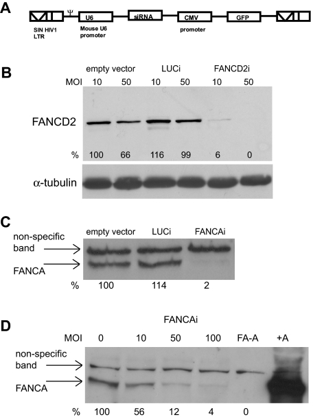
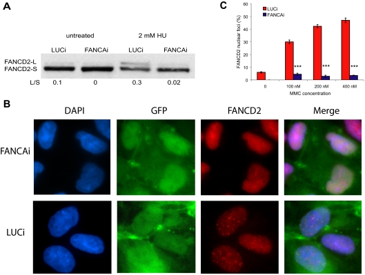
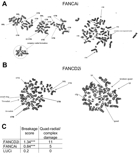
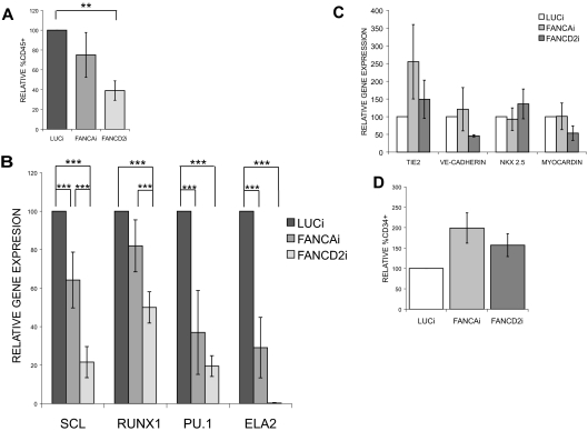
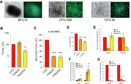
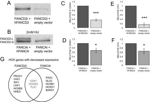
Fanconi anemia (FA) is a genetically heterogeneous, autosomal recessive disorder characterized by pediatric bone marrow failure and congenital anomalies. The effect of FA gene deficiency on hematopoietic development in utero remains poorly described as mouse models of FA do not develop hematopoietic failure and such studies cannot be performed on patients. We have created a human-specific in vitro system to study early hematopoietic development in FA using a lentiviral RNA interference (RNAi) strategy in human embryonic stem cells (hESCs). We show that knockdown of FANCA and FANCD2 in hESCs leads to a reduction in hematopoietic fates and progenitor numbers that can be rescued by FA gene complementation. Our data indicate that hematopoiesis is impaired in FA from the earliest stages of development, suggesting that deficiencies in embryonic hematopoiesis may underlie the progression to bone marrow failure in FA. This work illustrates how hESCs can provide unique insights into human development and further our understanding of genetic disease.


Figures

Figure 1
Figure 1
Efficient knockdown of FA proteins in hESCs by RNAi. (A) Schematic of the lentiviral vector used to deliver small interfering RNAs. (B) FANCD2 Western blot on lysates from GFP+ FACS for hESCs infected at MOI 10 and 50 with empty vector, LUCi, and FANCD2i virus at day 4 after infection. Percentage of knockdown is calculated using ImageJ software (National Institutes of Health) after normalization to α-tubulin levels. (C-D) FANCA Western blot on lysates from GFP+ FACS of hESCs infected either with empty vector, LUCi, and FANCAi virus at MOI 50 (C) or increasing MOIs of FANCAi virus (D) at day 4 after infection. Percentage of knockdown is calculated using ImageJ software after normalization to a nonspecific band. (D lane 5, FA-A) A lysate from a FA-A patient fibroblast; (lane 6, +A) the patient fibroblast line retrovirally corrected with FANCA.
Figure 2
Figure 2
FANCA knockdown results in marked reduction of FANCD2 monoubiquitination and nuclear foci formation. (A) FANCAi and LUCi hESCs were treated with 2mM HU for 24 hours, and lysates were subjected to Western blotting for FANCD2. FANCD2-L is the active, monoubiquitinated form of FANCD2-S. The ratio of L/S was calculated using ImageJ software. (B) FANCD2 immunofluorescence of FANCAi and LUCi hESCs treated with 200nM MMC treatment for 24 hours. DAPI (4,6 diamidino-2-phenylindole) stains for DNA content and GFP marks infected cells. (C) Quantification of FANCD2 nuclear foci after 100, 200, and 400 nM MMC treatment for 24 hours. Percentage FANCD2 nuclear foci (y-axis) represents the fraction of cells with 4 or more nuclear foci, as assayed by FANCD2 immunofluorescence. Data are represented as the average ± SEM; each data point represents 3 independent sets of 50 cells. ***P < .01 by Student t test.
Figure 3
Figure 3
FANCD2i and FANCAi hESCs exhibit increased chromosomal breakage and radial formation upon MMC treatment. (A-B) Representative metaphase spreads from FANCAi and FANCD2i hESCs after treatment with 200nM MMC for 24 hours. DEL indicates deletion; CTB, chromatid break; CTG, chromatid gap; and CSB, chromosome break. (C) Quantification of total damage/cell (additional details on breakage score calculation in “Chromosomal breakage analysis”) and quadriradials/complex damage (ring structures, extensive chromosomal breakage/rearrangement precluding precise quantitation) for 50 scored metaphases. ***P < .01 by χ2 test.
Figure 4
Figure 4
FANCAi and FANCD2i hESCs display reduced hematopoietic gene expression during in vitro differentiation. All data are presented as mean ± SEM. The number of biologic replicates (n) is reported for each panel. ***P < .01, **.01 < P < .03. (A) FACS analysis of day 20 EB-derived cells for the percentage of CD45+ cells. n = 8. (B-C) qRT-PCR analysis of representative hematopoietic (B), endothelial, and cardiac (C) markers in day 20 EBs. Relative gene expression (y-axis) is expressed as percentage and was calculated relative to LUCi expression levels after normalization with GAPDH. n = 6. (D) FACS analysis of day 20 EB-derived cells for the percentage of CD34+ cells. n = 6.
Figure 5
Figure 5
FANCD2i hESCs demonstrate significant reductions in hematopoietic CFUs. (A) Representative light and fluorescence micrographs of hematopoietic colonies. BFU-E indicates blast-forming unit erythroid; CFU-GM, colony-forming unit granulocyte/monocyte; and CFU-M, colony-forming unit macrophage. (B-C) Total hematopoietic CFUs from day 20 EB-derived cells plated in H4434 media (StemCell Technologies) without (B) or with (C) 5nM MMC and scored at day 14 after plating. Total CFUs are normalized directly to LUCi (B) or first normalized to baseline untreated total CFUs to assess MMC hypersensitivity of hematopoietic progenitors (C). Data are presented as mean ± SEM. n = 6, 3. (D) Normalized ratio of gamma-globin expression to epsilon-globin expression by qRT-PCR from CD45+ population of day 20 EB-derived cells. Data are presented as mean ± SEM. n = 6. (E-G) qRT-PCR analysis of gene expression from CD45+ population of day 20 EBs. Expression is relative to LUCi expression levels after normalization with GAPDH. Data are presented as mean ± SEM and n = 6. ***P < .01; **.01 < P < .03; *.03 < P < .05.
Figure 6
Figure 6
FA gene complementation rescues hematopoietic deficits. (A) FANCD2 Western blot on lysates from FANCD2i hESCs infected with pMMP-puro hFANCD2 or pMMP-puro (empty vector). (B) FANCAi hESCs infected with pMMP-puro hFANCA or pMMP-puro were treated with 2mM HU for 24 hours and lysates were subjected to Western blotting for FANCD2. FANCD2-L is the active, monoubiquitinated form of FANCD2-S. (C-D) FACS analysis of day 20 EB-derived cells for the percentage of CD45+ cells. Percentage CD45 is normalized to FANCD2i + hFANCD2 (C) or FANCAi + hFANCAi (D) and presented as ± SEM. n = 4, 6. (E-F) Total hematopoietic CFUs from day 20 EB-derived cells plated in H4434 media (StemCell Technologies) scored at day 14 after plating. Total CFUs are normalized to FANCD2i + hFANCD2 (E) or FANCAi + hFANCAi (F) and presented as ± SEM. n = 4, 6. (G) Venn diagram showing HOX genes with decreased expression in day 20 EBs. Eighty-four HOX genes were analyzed by quantitative PCR array (SABiosciences) and the top 8 genes, all with fold reduction greater than −1.5 relative to gene complement control, were included. FANCD2i + empty vector was compared with FANCD2I + hFANCD2 and FANCAi + empty vector was compared with FANCAi + hFANCA to generate the list of genes. Data are provided in supplemental Table 3. ***P < .01; *.03 < P < .05.

Comment in

References

    1. D'Andrea AD, Grompe M. The Fanconi anaemia/BRCA pathway. Nat Rev Cancer. 2003;3(1):23–34. - PubMed
    1. Tischkowitz M, Dokal I. Fanconi anaemia and leukaemia: clinical and molecular aspects. Br J Haematol. 2004;126(2):176–191. - PubMed
    1. Auerbach AD, Liu Q, Ghosh R, Pollack MS, Douglas GW, Broxmeyer HE. Prenatal identification of potential donors for umbilical cord blood transplantation for Fanconi anemia. Transfusion. 1990;30(8):682–687. - PubMed
    1. Kelly PF, Radtke S, von Kalle C, et al. Stem cell collection and gene transfer in Fanconi anemia. Mol Ther. 2007;15(1):211–219. - PubMed
    1. Alter BP. Arms and the man or hands and the child: congenital anomalies and hematologic syndromes. J Pediatr Hematol Oncol. 1997;19(4):287–291. - PubMed

Publication types

MeSH terms

Substances