. 2010 Jan 21;362(3):206-16.
doi: 10.1056/NEJMoa0900158.

Lethal skeletal dysplasia in mice and humans lacking the golgin GMAP-210

Affiliations

Lethal skeletal dysplasia in mice and humans lacking the golgin GMAP-210

Patrick Smits et al. N Engl J Med. .

Abstract

Background: Establishing the genetic basis of phenotypes such as skeletal dysplasia in model organisms can provide insights into biologic processes and their role in human disease.

Methods: We screened mutagenized mice and observed a neonatal lethal skeletal dysplasia with an autosomal recessive pattern of inheritance. Through genetic mapping and positional cloning, we identified the causative mutation.

Results: Affected mice had a nonsense mutation in the thyroid hormone receptor interactor 11 gene (Trip11), which encodes the Golgi microtubule-associated protein 210 (GMAP-210); the affected mice lacked this protein. Golgi architecture was disturbed in multiple tissues, including cartilage. Skeletal development was severely impaired, with chondrocytes showing swelling and stress in the endoplasmic reticulum, abnormal cellular differentiation, and increased cell death. Golgi-mediated glycosylation events were altered in fibroblasts and chondrocytes lacking GMAP-210, and these chondrocytes had intracellular accumulation of perlecan, an extracellular matrix protein, but not of type II collagen or aggrecan, two other extracellular matrix proteins. The similarities between the skeletal and cellular phenotypes in these mice and those in patients with achondrogenesis type 1A, a neonatal lethal form of skeletal dysplasia in humans, suggested that achondrogenesis type 1A may be caused by GMAP-210 deficiency. Sequence analysis revealed loss-of-function mutations in the 10 unrelated patients with achondrogenesis type 1A whom we studied.

Conclusions: GMAP-210 is required for the efficient glycosylation and cellular transport of multiple proteins. The identification of a mutation affecting GMAP-210 in mice, and then in humans, as the cause of a lethal skeletal dysplasia underscores the value of screening for abnormal phenotypes in model organisms and identifying the causative mutations.


Conflict of interest statement

No potential conflict of interest relevant to this article was reported.

Figures

Figure 1
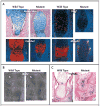
Figure 1. Effects of Trip11 Mutation in Mice
Panel A shows a wild-type fetal mouse and a fetal mouse with an induced nonsense mutation in Trip11, the gene encoding GMAP-210, on embryonic day 18.5. The mouse with the mutation has a domed skull, short snout, short trunk, short limbs, and omphalocele (arrowhead). In Panel B (top), the staining of cartilage with Alcian blue and bone with alizarin red on embryonic day 17.5 in a wild-type mouse and a mouse with the Trip11 mutation reveals the absence of mineralization in the skull (arrowhead), rib cage, and limbs (arrows) in the mutant. In Panel B, bottom, staining of cartilage and bone in the vertebral columns of newborn wild-type and mutant mice reveals the absence of mineralization in the vertebral body of the mutant (arrow). In Panel C, immunoblotting of GMAP-210 in immunoprecipitated samples of primary skin fibroblasts from a wild-type mouse, a heterozygous mouse, and a mutant mouse shows that GMAP-210 is not detectable in the mutant fibroblasts. IgG, which was used in the immunoprecipitation reaction, also serves as a loading control. In Panel D, in situ hybridization of specimens of wild-type shoulder girdle and humerus obtained on embryonic day 15.5, with the use of Trip11 antisense and sense RNA probes, shows the expression of Trip11 in all cell types, with an absence of increased expression in cartilage (arrow) and bone (arrowhead). Red indicates Trip11 mRNA expression. The nuclei of cells have been stained with 4′,6-diamidino-2-phenylindole, which fluoresces blue. Panel E shows histologic sections of the shoulder girdle and humerus from wild-type and mutant mice, obtained on embryonic day 15.5 and stained with nuclear-fast red and Alcian blue. The cartilage in the mutant humerus shows less staining with Alcian blue and contains chondrocytes with increased cell sizes. The mutant specimen also lacks an organized growth zone of flattened chondrocytes and a primary ossification (PO) center, although it does have a bone collar (arrow). Higher magnification images from the wild-type and mutant growth zones show the increased size of chondrocytes and the lack of chondrocyte columnar organization in the mutant cartilage.
Figure 2
Figure 2. Effects of GMAP-210 Deficiency on Hypertrophic Differentiation, Proliferation, and Apoptosis of Chondrocytes in Mice
Panel A shows sections through the primary ossification centers of humeri from wild-type and mutant mice that were obtained on embryonic day 14.5, stained with Alcian blue, and subjected to in situ hybridization. Col10a1 expression has not separated into two domains in the mutant humerus, and there is no cartilage expression of the late-stage hypertrophic chondrocyte markers vascular endothelial growth factor (Vegf) and matrix metalloproteinase 13 (Mmp13). Mmp13 is expressed only by periosteal osteoclasts in the mutant humerus (arrowheads). Sections through shoulder joints and humeri obtained on embryonic day 15.5 show that there is no difference in the incorporation of bromodeoxyuridine (BrdU) between wild-type and mutant chondrocytes adjacent to the joint (Panel B, arrowheads) but that in the mutant cartilage there is no other area of BrdU incorporation. Use of the terminal deoxynucleotidyl transferase dUTP biotin nick end labeling (TUNEL) assay on histologic sections of elbow joints obtained on embryonic day 17.5 show that the mutant humerus (H) and ulna (U) have many TUNEL-positive cells, which appear blue in the sections, whereas the wild-type humerus and ulna do not (Panel C).
Figure 3
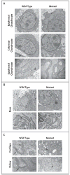
Figure 3. Swollen Endoplasmic Reticulum Cisternae and Abnormal Golgi Architecture in Murine Chondrocytes Lacking GMAP-210
Electron micrographs of chondrocytes from wild-type and mutant humeri obtained on embryonic day 15.5 show increased swelling of the endoplasmic reticulum in epiphyseal and columnar chondrocytes in mice lacking GMAP-210 (Panel A), as compared with their wild-type counterparts. A higher-magnification view of the epiphyseal chondrocytes showing ribosomes along the membrane surface of swollen cisternae in the mutant indicates that this is part of the endoplasmic reticulum. Electron micrographs of osteoblasts from the bone collar of wild-type and mutant humeri (Panel B, top row, and at higher magnification in the bottom row) show increased swelling of the endoplasmic reticulum in the mutant osteoblast. Panel C shows Golgi stacks in wild-type cartilage cells obtained on embryonic day 13.5 (top row, arrows) and wild-type kidney cells obtained on embryonic day 15.5 (bottom row, arrow) but not in mutant cells. The Golgi apparatus in the mutant cells consists of a collection of vesicles and enlarged cisternae (arrowheads).
Figure 4
Figure 4. Impaired Post-Translational Protein Processing and Secretion in Murine Cells Lacking GMAP-210
Panel A shows immunofluorescence staining for GM130, a cis-Golgi protein, and Arl1, a trans-Golgi protein, in wild-type and mutant primary skin fibroblasts. The immunofluorescence staining shows that mutant fibroblasts lack the reticular pattern that is present in wild-type fibroblasts and instead have small vesicles that contain these Golgi-associated proteins. The gray scale is used to indicate fluorescence in the images for which only one protein is shown, and color is used for the merged images in which both proteins are shown. The merged images show that the structures containing GM130 (red) and Arl1 (green) do not overlap in wild-type or in mutant fibroblasts. Although the appearance of the Golgi apparatus differs between mutant fibroblasts and wild-type fibroblasts, the lack of overlap between GM130-containing vesicles and Arl1-containing vesicles indicates that mutant fibroblasts maintain the cis-trans polarity of the Golgi apparatus. Panels B and C show the results of a transport assay performed with vesicular stomatitis viral G protein fused to green fluorescence protein (VSVGts–GFP) in wild-type and mutant primary skin fibroblasts. In Panel B, fluorescence localization of VSVGts–GFP and immunofluorescence localization of GM130 was performed in wild-type and mutant primary skin fibroblasts after the temperature of the infected cells had been shifted from 40°C to 32°C for 20 minutes. The gray scale is used to indicate fluorescence in the images for which only one protein is shown, and color is used for the merged images in which both proteins are shown. The merged images show that VSVGts–GFP (green) and GM130 (red) have substantial overlap (yellow) in wild-type and in mutant cells, indicating that transport from the endoplasmic reticulum to the Golgi apparatus is not blocked in mutant cells. Panel C shows a Western blot analysis of a representative VSVGts–GFP transport assay and a graph depicting the means (±SD) of three independent experiments using primary skin fibroblasts from mice that were heterozygous or homozygous for the Trip11 mutation. After 1 hour and 3 hours at 32°C, cells that were completely lacking GMAP-210 (homozygous) had a lower mean ratio of endoglycosidase H–insensitive (endoH-i) VSVGts–GFP to endoglycosidase H–sensitive (EndoH-s) VSVGts–GFP than heterozygous cells; this finding is consistent with the view that the efficiency of Golgi glycosylation is reduced in fibroblasts with mutant GMAP-210. In Panel D, electron micrographs of extracellular matrix from epiphyseal cartilage in wild-type and mutant humeri, obtained on embryonic day 17.5, show that the density of collagen fibrils is similar but that the glycosaminoglycan-containing complexes, which normally collapse and appear granular after the electron-microscopical fixation process (arrows), are smaller in the mutant extracellular matrix. Panel E shows changes in the distribution of perlecan (which can be seen with immunofluorescence detection and are depicted in the gray scale) between histologic sections of wild-type and mutant humeri obtained on embryonic day 15.5 (top row). At higher magnification, immunofluorescence detection of perlecan (red) and the endoplasmic reticulum protein calreticulin (green) shows that mutant chondrocytes have greater retention of perlecan within their endoplasmic reticulum than do wild-type chondrocytes, as indicated by the stronger intracellular fluorescence and partial overlap (yellow) with calreticulin in the merged image. In Panel F, immunofluorescence detection of perlecan in the skin of wild-type and mutant mice on embryonic day 15.5 shows an accumulation of perlecan in the mutant dermal fibroblasts (arrowheads). The arrows indicate the epidermal basement membrane; E denotes epidermis, and D dermis.
Figure 5
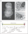
Figure 5. Mutations in TRIP11 and Human Achondrogenesis Type 1A
In Panel A, a radiograph of a 27-week-old human fetus with a diagnosis of achondrogenesis type 1A reveals a lack of mineralization in the skull and the vertebral column (arrows) and short limbs. The electron micrographs at the right show chondrocytes from two unrelated fetuses with achondrogenesis type 1A, with swollen endoplasmic reticulum (arrows) and vesicular Golgi apparatus (arrowhead in upper image). In Panel B, the structure of the GMAP-210 protein is shown with the relative location and sizes of its 21 coding exons indicated below. ALPS (amphipathic lipid sensor domain) and GRAB (GRIP-related Arf-binding domain) reportedly mediate interactions between GMAP-210 and membranes in vitro. In the diagram below the protein, arrows point from the mutations that were identified in 10 unrelated patients with achondrogenesis type 1A to their approximate locations within the exons. Two mutations (c.202-2A→G and c.589-2A→G) affect intronic splice-acceptor sites. Five mutations (p.R264X, p.R1028X, p.Q1160X, p.R1167X, and p.W1224X) are nonsense mutations. The remaining mutations are frameshift mutations. Several patients, including those from consanguineous unions, were homozygous for mutations, whereas others were compound heterozygotes. Some mutations were found in more than one patient.

Comment in

Similar articles

Cited by

References

    1. Barr FA, Short B. Golgins in the structure and dynamics of the Golgi apparatus. Curr Opin Cell Biol. 2003;15:405–13. - PubMed
    1. Sztul E, Lupashin V. Role of tethering factors in secretory membrane traffic. Am J Physiol Cell Physiol. 2006;290:C11–C26. - PubMed
    1. Molz G, Spycher MA. Achondrogenesis type I: light and electron-microscopic studies. Eur J Pediatr. 1980;134:69–74. - PubMed
    1. Herron BJ, Lu W, Rao C, et al. Efficient generation and mapping of recessive developmental mutations using ENU mutagenesis. Nat Genet. 2002;30:185–9. - PubMed
    1. Moran JL, Bolton AD, Tran PV, et al. Utilization of a whole genome SNP panel for efficient genetic mapping in the mouse. Genome Res. 2006;16:436–40. - PMC - PubMed

Publication types

MeSH terms