Transcriptional role of cyclin D1 in development revealed by a genetic-proteomic screen
- PMID: 20090754
- PMCID: PMC2943587
- DOI: 10.1038/nature08684
Transcriptional role of cyclin D1 in development revealed by a genetic-proteomic screen
Abstract
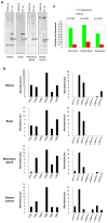
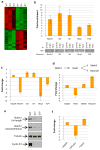
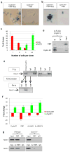
Cyclin D1 belongs to the core cell cycle machinery, and it is frequently overexpressed in human cancers. The full repertoire of cyclin D1 functions in normal development and oncogenesis is unclear at present. Here we developed Flag- and haemagglutinin-tagged cyclin D1 knock-in mouse strains that allowed a high-throughput mass spectrometry approach to search for cyclin D1-binding proteins in different mouse organs. In addition to cell cycle partners, we observed several proteins involved in transcription. Genome-wide location analyses (chromatin immunoprecipitation coupled to DNA microarray; ChIP-chip) showed that during mouse development cyclin D1 occupies promoters of abundantly expressed genes. In particular, we found that in developing mouse retinas-an organ that critically requires cyclin D1 function-cyclin D1 binds the upstream regulatory region of the Notch1 gene, where it serves to recruit CREB binding protein (CBP) histone acetyltransferase. Genetic ablation of cyclin D1 resulted in decreased CBP recruitment, decreased histone acetylation of the Notch1 promoter region, and led to decreased levels of the Notch1 transcript and protein in cyclin D1-null (Ccnd1(-/-)) retinas. Transduction of an activated allele of Notch1 into Ccnd1(-/-) retinas increased proliferation of retinal progenitor cells, indicating that upregulation of Notch1 signalling alleviates the phenotype of cyclin D1-deficiency. These studies show that in addition to its well-established cell cycle roles, cyclin D1 has an in vivo transcriptional function in mouse development. Our approach, which we term 'genetic-proteomic', can be used to study the in vivo function of essentially any protein.
Figures


References
-
- Malumbres M, Barbacid M. Cell cycle, CDKs and cancer: a changing paradigm. Nat Rev Cancer. 2009;9:153–66. - PubMed
-
- Sherr CJ, Roberts JM. CDK inhibitors: positive and negative regulators of G1-phase progression. Genes Dev. 1999;13:1501–12. - PubMed
-
- Fantl V, Stamp G, Andrews A, Rosewell I, Dickson C. Mice lacking cyclin D1 are small and show defects in eye and mammary gland development. Genes Dev. 1995;9:2364–72. - PubMed
-
- Sicinski P, et al. Cyclin D1 provides a link between development and oncogenesis in the retina and breast. Cell. 1995;82:621–30. - PubMed
-
- Yu Q, Geng Y, Sicinski P. Specific protection against breast cancers by cyclin D1 ablation. Nature. 2001;411:1017–21. - PubMed
Publication types
MeSH terms
Substances
Associated data
- Actions
Grants and funding
- HG004069/HG/NHGRI NIH HHS/United States
- P01 CA109901/CA/NCI NIH HHS/United States
- P01 CA080111/CA/NCI NIH HHS/United States
- R01 EYO9676/PHS HHS/United States
- R01 EY009676/EY/NEI NIH HHS/United States
- HG3456/HG/NHGRI NIH HHS/United States
- R01 HG004069/HG/NHGRI NIH HHS/United States
- R01 HG003456/HG/NHGRI NIH HHS/United States
- A15603/CRUK_/Cancer Research UK/United Kingdom
- R01 CA108420/CA/NCI NIH HHS/United States
- 15603/CRUK_/Cancer Research UK/United Kingdom
- R01 HG002668/HG/NHGRI NIH HHS/United States
- R01 EY008064/EY/NEI NIH HHS/United States
LinkOut - more resources
Full Text Sources
Other Literature Sources
Molecular Biology Databases
Research Materials

