

Analysis of k-ras nuclear expression in fibroblasts and mesangial cells
- PMID: 20090846
- PMCID: PMC2806826
- DOI: 10.1371/journal.pone.0008703
Analysis of k-ras nuclear expression in fibroblasts and mesangial cells
Abstract
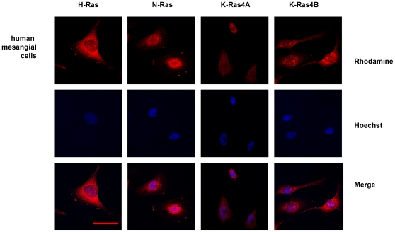
Background: Ras GTPases are considered cytoplasmic proteins that must be localized to cell membranes for activation, and there are few evidences of the presence of any Ras isoform in nuclei of eukaryotic cells.
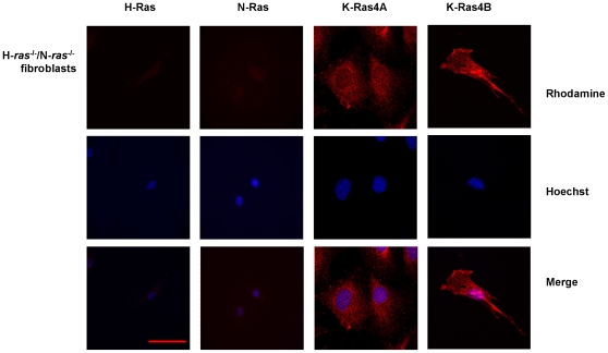
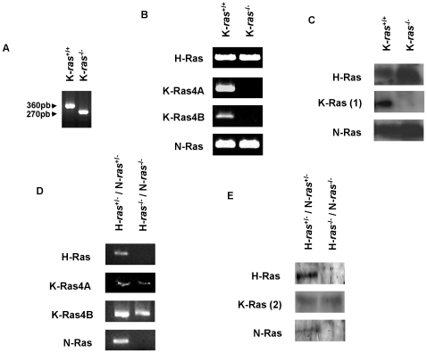
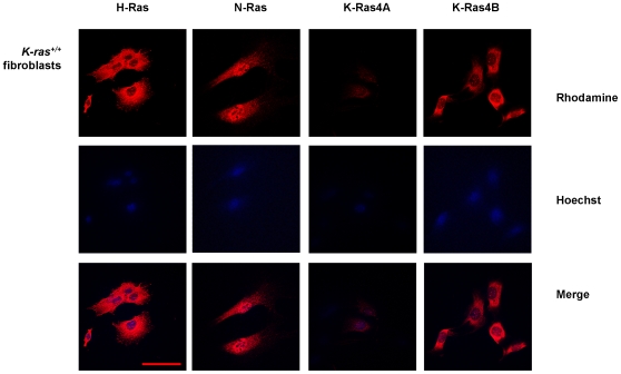
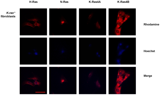
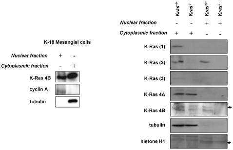
Methodology/principal findings: Using conventional antibodies and inmunocytochemistry, differential centrifugation and western blot, we have observed the putative presence of K-Ras isoform in the nuclei of fibroblasts and mesangial cells. In order to avoid cross-reactions with other Ras isoforms, and using antibodies against K-Ras (R-3400, H3845-M01, sc-30) or pan-Ras (05-516, OP40) in cells that only expressed the K-Ras isoform (fibroblasts obtained from H-ras(-/-),N-ras(-/-) mice) we also detected some nuclear positive expression. To further probe the identity of nuclear K-Ras, we have generated K-Ras knockout (K-ras(-/-)) embrionary fibroblasts by mating of K-ras(+/-) heterozygote mice. Using specific antibodies, only H- and N-Ras isoforms were observed in the cytoplasm of K-ras(-/-) fibroblasts. However, both K-Ras4A and K-Ras4B positive signals were detected by immunocytochemistry and Western blot with two commercial antibodies (sc-522 and sc-521 against each isoforms, respectively) in both cytoplasm and nuclei from K-ras(-/-) fibroblasts.
Conclusions/significance: We show that the presence of K-Ras4B in fibroblast nuclei, already described by other authors, is probably due to a cross-reaction of the antibody with an undetermined nucleolar protein. Although this study also shows the possible nuclear expression of K-Ras isoform in fibroblasts or in mesangial cells, it also reveals the importance of being cautious in these studies about distribution of protein isoforms due to some important limitations imposed by the unspecificity of the antibodies or contaminations in cellular preparations.
Conflict of interest statement
Figures
Similar articles
-
Expression of Ras GTPases in normal kidney and in glomerulonephritis.Nephrol Dial Transplant. 2003 Nov;18(11):2284-92. doi: 10.1093/ndt/gfg375. Nephrol Dial Transplant. 2003. PMID: 14551355
-
Alternative splicing of the K-ras gene in mouse tissues and cell lines.Exp Lung Res. 2001 Apr-May;27(3):255-67. doi: 10.1080/019021401300054028. Exp Lung Res. 2001. PMID: 11293328
-
The K-Ras 4A isoform promotes apoptosis but does not affect either lifespan or spontaneous tumor incidence in aging mice.Exp Cell Res. 2006 Jan 1;312(1):16-26. doi: 10.1016/j.yexcr.2005.10.004. Epub 2005 Nov 4. Exp Cell Res. 2006. PMID: 16271715
-
A New View of Ras Isoforms in Cancers.Cancer Res. 2016 Jan 1;76(1):18-23. doi: 10.1158/0008-5472.CAN-15-1536. Epub 2015 Dec 10. Cancer Res. 2016. PMID: 26659836 Free PMC article. Review.
-
Translocational inefficiency of intracellular proteins in senescence of human diploid fibroblasts.Ann N Y Acad Sci. 2001 Apr;928:176-81. doi: 10.1111/j.1749-6632.2001.tb05647.x. Ann N Y Acad Sci. 2001. PMID: 11795508 Review.
Cited by
-
Post-embryonic endogenous expression and localization of LET-60/Ras in C. elegans.MicroPubl Biol. 2023 Aug 25;2023:10.17912/micropub.biology.000931. doi: 10.17912/micropub.biology.000931. eCollection 2023. MicroPubl Biol. 2023. PMID: 37692087 Free PMC article.
-
Analysis of K-Ras Interactions by Biotin Ligase Tagging.Cancer Genomics Proteomics. 2017 Jul-Aug;14(4):225-239. doi: 10.21873/cgp.20034. Cancer Genomics Proteomics. 2017. PMID: 28647697 Free PMC article.
-
Targeted genetic and small molecule disruption of N-Ras CaaX cleavage alters its localization and oncogenic potential.Bioorg Chem. 2024 Jun;147:107316. doi: 10.1016/j.bioorg.2024.107316. Epub 2024 Mar 27. Bioorg Chem. 2024. PMID: 38583246 Free PMC article.
-
Stress-Induced Phosphorylation of Nuclear YB-1 Depends on Nuclear Trafficking of p90 Ribosomal S6 Kinase.Int J Mol Sci. 2018 Aug 18;19(8):2441. doi: 10.3390/ijms19082441. Int J Mol Sci. 2018. PMID: 30126195 Free PMC article.
-
Phenotypic Switching of Naïve T Cells to Immune-Suppressive Treg-Like Cells by Mutant KRAS.J Clin Med. 2019 Oct 18;8(10):1726. doi: 10.3390/jcm8101726. J Clin Med. 2019. PMID: 31635338 Free PMC article.
References
-
- Ellis CA, Clark G. The importance of being K-Ras. Cell Signal. 2000;12:425–434. - PubMed
-
- Takai Y, Sasaki T, Matozaki T. Small GTP-binding proteins. Physiol Rev. 2001;81:153–208. - PubMed
-
- Barbacid M. ras genes. Annu Rev Biochem. 1987;56:779–827. - PubMed
-
- Dhanasekaran N, Premkumar Reddy E. Signaling by dual specificity kinases. Oncogene. 1998;17:1447–1455. - PubMed
-
- Kerkhoff E, Rapp UR. Cell cycle targets of Ras/Raf signalling. Oncogene. 1998;17:1457–1462. - PubMed
Publication types
MeSH terms
Substances
LinkOut - more resources
Full Text Sources
Molecular Biology Databases
Research Materials
Miscellaneous
