. 2010 Jan 14;5(1):e8704.
doi: 10.1371/journal.pone.0008704.

A proteomic approach for comprehensively screening substrates of protein kinases such as Rho-kinase

Affiliations

A proteomic approach for comprehensively screening substrates of protein kinases such as Rho-kinase

Mutsuki Amano et al. PLoS One. .

Abstract

Background: Protein kinases are major components of signal transduction pathways in multiple cellular processes. Kinases directly interact with and phosphorylate downstream substrates, thus modulating their functions. Despite the importance of identifying substrates in order to more fully understand the signaling network of respective kinases, efficient methods to search for substrates remain poorly explored.

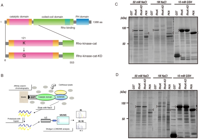
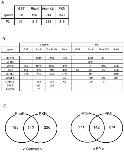
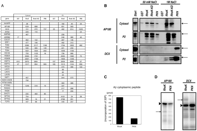
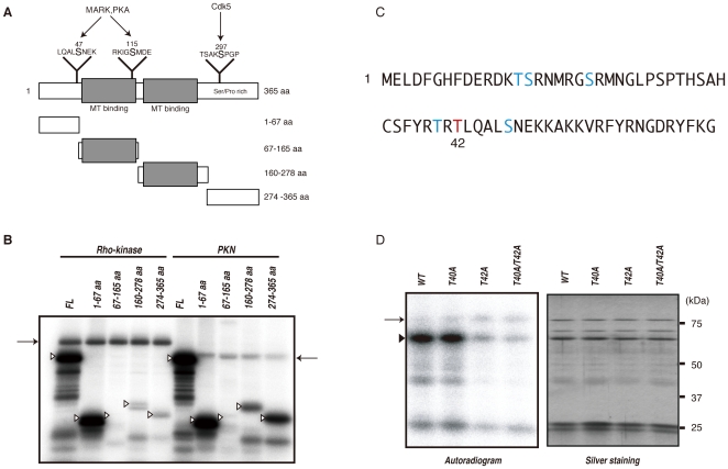
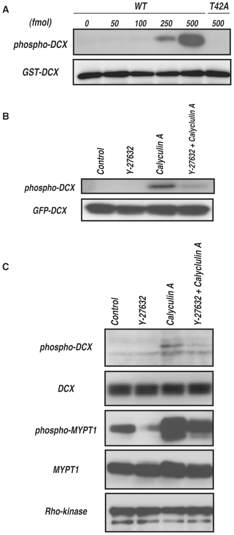
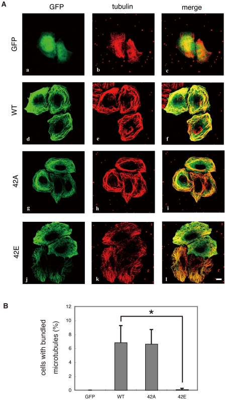
Methodology/principal findings: We combined mass spectrometry and affinity column chromatography of the catalytic domain of protein kinases to screen potential substrates. Using the active catalytic fragment of Rho-kinase/ROCK/ROK as the model bait, we obtained about 300 interacting proteins from the rat brain cytosol fraction, which included the proteins previously reported as Rho-kinase substrates. Several novel interacting proteins, including doublecortin, were phosphorylated by Rho-kinase both in vitro and in vivo.

Conclusions/significance: This method would enable identification of novel specific substrates for kinases such as Rho-kinase with high sensitivity.


Conflict of interest statement

Competing Interests: The authors have declared that no competing interests exist.

Figures

Figure 1
Figure 1. Isolation of interacting proteins for the catalytic domain of Rho-kinase.
(A) Domain structure of Rho-kinase and the constructs used for affinity column chromatography. (B) Strategy for isolation of protein kinase substrates. (C, D) Isolation of Rho-kinase-cat-interacting proteins from rat brain cytosol (C) and P2 (D) fractions. The cytosolic or P2 fraction of rat brain lysate was loaded onto a Glutathione-Sepharose column coated with either GST, GST-Rho-kinase-cat, GST-Rho-kinase-cat-KD or GST-PKN-cat. The bound proteins were eluted by addition of 1 M NaCl after washing with 50 mM NaCl. The eluates were analyze by SDS-PAGE, and visualized by silver staining. Arrowheads indicate the GST-tagged proteins used as baits.
Figure 2
Figure 2. Identification of Rho-kinase-cat-interacting proteins.
(A) Total numbers of hit proteins detected in eluates off each affinity column. (B) Known substrates for Rho-kinase detected in eluates off each affinity column. The numbers indicate protein scores by Mascot analysis. (C) The numbers of proteins specifically or commonly detected in GST-Rho-kinase-cat and GST-PKN-cat columns are shown. These results are representatives of at least three independent experiments.
Figure 3
Figure 3. Identification of novel substrates for Rho-kinase.
(A) Protein scores of APP, AP180, and DCX in eluates off each affinity column. (B) Immunoblot analysis of eluates with anti-AP180 and -DCX Abs. Eluates off affinity columns with 50 mM and 1 M NaCl were subjected to immunoblot analysis with anti-AP180 and -DCX Abs. AP180 was detected in both 50 mM and 1 M NaCl eluates off the Rho-kinase-cat column, but was barely detectable in eluates off the PKN-cat column. On the contrary, DCX was strongly detected in eluates off the PKN-cat column, and moderately off the Rho-kinase-cat column. Arrows indicate the positions of AP180 and DCX. The lower bands are supposed to be degradation products or splice variants. (C) The APP cytoplasmic peptide was incubated with GST-Rho-kinase-cat or GST-PKN-cat in the presence of 100 µM [γ-32P] ATP for 1 h at 30°C. The reaction mixtures were applied to P81 paper and subjected to scintillation counting. (D) GST-AP180 (left) or GST-DCX (right) was incubated with GST-Rho-kinase-cat or GST-PKN-cat in the presence of 100 µM [γ-32P] ATP for 1 h at 30°C. The reaction mixtures were subjected to SDS-PAGE, and phosphorylated proteins were imaged by autoradiography. Arrowheads and arrows indicate substrates and autophosphorylation of Rho-kinase, respectively. These results are representatives of at least three independent experiments.
Figure 4
Figure 4. Identification of phosphorylation sites of DCX by Rho-kinase.
(A) Schematic representation of the domain structures and deletion mutants of DCX. Sites phosphorylated by MARK, PKA and Cdk5 are also shown. (B) Phosphorylation of DCX deletion mutants. The indicated GST-DCX fragments were phosphorylated by GST-Rho-kinase-cat or GST-PKN-cat. The phosphorylated proteins were imaged by autoradiography. Open arrowheads and arrows indicate the positions of substrates and autophosphorylation of kinases, respectively. These results are representatives of at least three independent experiments. (C) Sequence of and potential phosphorylation sites within the 1–67 aa region. The major phosphorylation site was identified as Thr42 (red). (D) Phosphorylation of DCX mutants with amino acid substitutions. GST-DCX-WT, -T40A, -T42A or -T40A/T42A was incubated with Rho-kinase-cat and 50 µM [γ-32P] ATP for 10 min at 30°C. The reaction mixtures were subjected to SDS-PAGE and GST-fused proteins were visualized by silver staining (right). Phosphorylated proteins were imaged by autoradiography (left). Arrowhead and arrow indicate the positions of substrates and autophosphorylation of kinase, respectively. These results are representatives of at least three independent experiments.
Figure 5
Figure 5. Phosphorylation of DCX in vivo.
(A) Specificity of the antibody against DCX phosphorylated at Thr-42 (anti-pT42 Ab). One pmol of GST-DCX containing the indicated amounts of phosphorylated GST-DCX-WT or -T42A was subjected to SDS-PAGE, followed by immunoblot analysis with anti-pT42 Ab (upper panel) or anti-GST Ab (lower panel). (B) Phosphorylation of DCX in COS7 cells. GFP-DCX was transiently expressed into COS7 cells. The transfected cells were treated with DMSO or 20 µM Y-27632 for 15 min, and then treated with or without 0.1 µM calyculin A for 10 min. The cell lysates were analyzed by immunoblot analysis with anti-pT42 Ab (upper panel) or anti-GFP Ab (lower panel). These results are representatives of at least three independent experiments. (C) Phosphorylation of DCX in hippocampal neurons at DIV1. The cells were treated with DMSO or 20 µM Y-27632 for 20 min, and then treated with or without 50 nM calyculin A for 7 min. The cell lysates were analyzed by immunoblot analysis with anti-pT42 Ab or anti-DCX Ab. Phosphorylation of MYPT1 was also examined with anti-MYPT1 pT853 Ab. These results are representatives of at least three independent experiments.
Figure 6
Figure 6. Effect of mutations of DCX at phosphorylation sites on microtubule organization in HeLa cells.
(A) Effect of overexpression of DCX-WT, -T42A and -T42E in HeLa cells. DCX mutants with substitutions at phosphorylation sites were expressed in HeLa cells. HeLa cells expressing GFP-DCX or its mutants were fixed in methanol for 10 min at room temperature. After washing, the cells were immunostained with anti-GFP and anti-tubulin Abs. Colors indicate GFP (green) and tubulin (red). These results are representatives of at least three independent experiments. Scale bar, 10 µm. (B) The percentages of cells with highly bundled microtubules. Data are means ± SD of three independent experiments. Asterisks indicate that there is a significant difference from the value of control cells (p<0.05).

References

    1. Ptacek J, Devgan G, Michaud G, Zhu H, Zhu X, et al. Global analysis of protein phosphorylation in yeast. Nature. 2005;438:679–684. - PubMed
    1. Schmelzle K, White FM. Phosphoproteomic approaches to elucidate cellular signaling networks. Curr Opin Biotechnol. 2006;17:406–414. - PubMed
    1. Tedford NC, Hall AB, Graham JR, Murphy CE, Gordon NF, et al. Quantitative analysis of cell signaling and drug action via mass spectrometry-based systems level phosphoproteomics. Proteomics. 2009;9:1469–1487. - PubMed
    1. Machida M, Kosako H, Shirakabe K, Kobayashi M, Ushiyama M, et al. Purification of phosphoproteins by immobilized metal affinity chromatography and its application to phosphoproteome analysis. Febs J. 2007;274:1576–1587. - PubMed
    1. Ubersax JA, Ferrell JE., Jr Mechanisms of specificity in protein phosphorylation. Nat Rev Mol Cell Biol. 2007;8:530–541. - PubMed

Publication types