. 2010 Jan 18;5(1):e8756.
doi: 10.1371/journal.pone.0008756.

Manipulating IL-2 availability amid presentation of donor MHC antigens suppresses murine alloimmune responses by inducing regulatory T cells

Affiliations

Manipulating IL-2 availability amid presentation of donor MHC antigens suppresses murine alloimmune responses by inducing regulatory T cells

Shuzi Zhang et al. PLoS One. .

Abstract

Background: Major histocompatibility complex (MHC) antigens are important for alloimmune responses as well as immune tolerance. Previous studies have shown that presentation of donor MHC antigens by donor-specific transfusion prior to or upon transplantation promotes transplant tolerance induced by other agents. However, it is unclear whether presentation of donor MHC antigens by DNA vaccination induces long-term allograft survival.

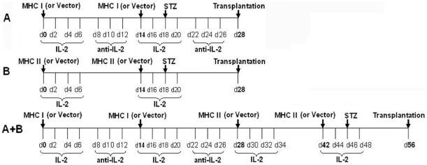
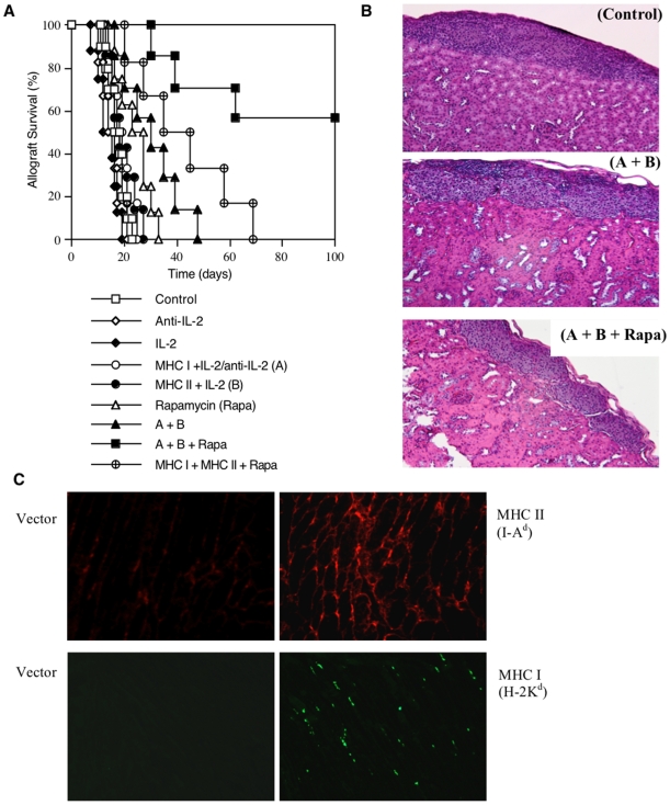
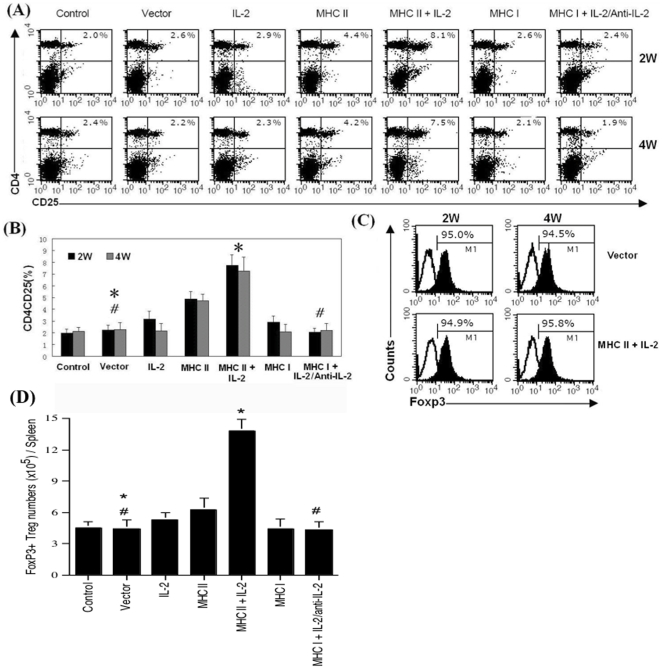
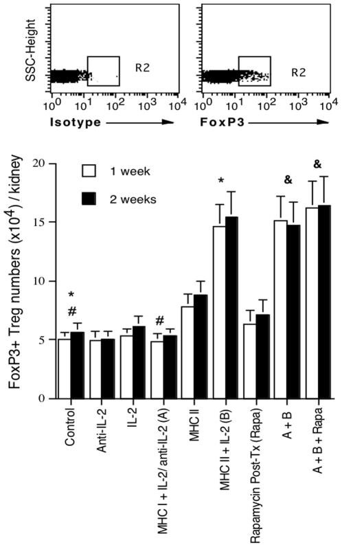
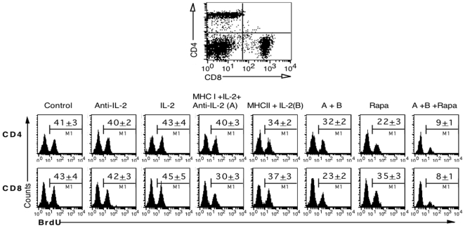
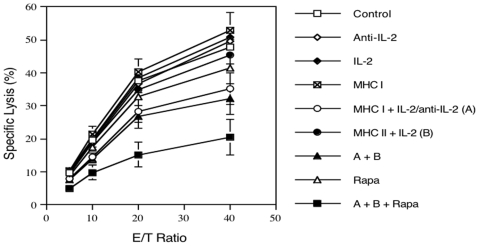
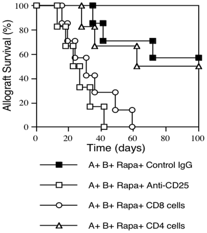
Methodology/principal findings: We investigated whether presentation of MHC class-II and/or class-I donor antigens by DNA vaccination suppresses alloimmune responses and promotes long-term allograft acceptance. We initially found that presentation of both MHC donor antigens by DNA vaccination itself prior to transplantation fails to significantly prolong islet allograft survival in otherwise untreated mice. However, islet allograft survival was significantly prolonged when MHC class-II DNA vaccination was accompanied with IL-2 administration (MHCII + IL-2) while MHC class-I DNA vaccination was followed by IL-2 and subsequent neutralizing anti-IL-2 treatments (MHCI + IL-2/anti-IL-2). Especially, this protocol promoted long-term allograft survival in the majority of recipients (57%) when combined with low doses of rapamycin post-transplantation. Importantly, MHCII + IL-2 induced FoxP3+ Treg cells in both spleens and grafts and suppressed graft-infiltrating CD4+ cell proliferation, whereas MHCI + IL-2/anti-IL-2 mainly inhibited graft-infiltrating CD8+ cell proliferation and donor-specific CTL activity. The combined protocol plus rapamycin treatment further reduced both CD4+ and CD8+ T cell proliferation as well as donor-specific CTL activity but spared FoxP3+ Treg cells. Depleting CD25+ Treg cells or adoptive transfer of pre-sensitized CD8+ T cells abolished this long-term allograft survival.

Conclusions/significance: Manipulating IL-2 availability during presentation of MHC class-II and class-I donor antigens by DNA vaccination pre-transplantation induces Treg cells, suppresses alloimmune responses and promotes long-term allograft survival.


Conflict of interest statement

Competing Interests: The authors have declared that no competing interests exist.

Figures

Figure 1
Figure 1. MHC immunization procedure.
Mice were immunized with donor MHC I and/or MHC II DNA vaccines and treated with recombinant IL-2 and/or anti-IL-2 Ab as well as streptozotocin (STZ) before islet transplantation.
Figure 2
Figure 2. Donor MHC DNA vaccination and administration of IL-2 regimen prior to transplantation promote long-term islet allograft survival.
Prior to transplantation, B6 mice were untreated (□, n = 10) or treated with neutralizing anti-IL-2 (◊, n = 6) alone, recombinant IL-2 alone (υ, n = 8), donor MHC class-I DNA vaccination plus both IL-2 and subsequent anti-IL-2 (MHC I + IL-2/anti-IL-2: A) (○, n = 6), donor MHC class-II DNA vaccination plus IL-2 (MHC II + IL-2: B) (λ, n = 7), or both MHC I and MHC II vaccination plus IL-2 regimen (A + B) (σ, n = 7). In some groups, rapamycin control mice (Rapa) (▵, n = 8) or mice treated with both MHC vaccinations and IL-2 regimen (A + B + Rapa) (ν, n = 7) or both vaccinations without IL-2 (MHC I + MHC II + Rapa) (⊕, n = 6) received low doses of rapamycin post-transplantation. (A). Islet allograft rejection was observed. (B). H&E staining on kidney sections at the time of rejection or 100 days after transplantation. (C). Immunofluorescence staining on muscular frozen sections for the expression of donor MHC I (H-2Kd) or MHC II (I-Ad) three days after donor MHC DNA vaccination in a recipient. One representative of three independent experiments is shown.
Figure 3
Figure 3. Donor MHC II DNA vaccination and administration of IL-2 prior to transplantation induce FoxP3+ Treg cells.
B6 mice were treated with recombinant IL-2 alone, anti-IL-2 alone, immunization with vector, MHC I or MHC II alone, or both IL-2 regimen and donor MHC DNA vaccination in recipients as indicated. Two and four weeks after secondary immunization, spleen cells were stained for CD4, CD8 and FoxP3 expression and analyzed by FACS. (A). Density plot is shown for the percentage of CD4+CD25+ Treg cells from one representative experiment. (B). Data are presented as mean ± SEM and represent an average of three independent experiments (*P<0.05 and #P>0.05). (C). Histograms shown are percentages of FoxP3+ cells after gating on CD4+CD25+ cells from control group. One representative of three experiments is shown. (D). The absolute numbers of FoxP3+ Treg cells per spleen two weeks after final immunization were also calculated (*P<0.05 and #P>0.05).
Figure 4
Figure 4. The absolute number of FoxP3-positive Treg cells in allografts.
Prior to transplantation, B6 mice were immunized with donor MHC DNA vaccines and/or treated with IL-2 regimen as described in Figure 1. One and two weeks after receiving Balb/C islets under the kidney capsule, recipient mice were sacrificed and graft-infiltrating cells were isolated and stained for FoxP3 to quantify FoxP3+ Treg cells by FACS. FoxP3+ cell numbers were calculated by the formula that total cell numbers per kidney time the percentage of FoxP3+ cells (%). Results are shown as mean ± SEM from four independent experiments (*P<0.05, #P>0.05, and &P>0.05).
Figure 5
Figure 5. In vivo proliferation of graft-infiltrating T cells.
Prior to transplantation, B6 mice were immunized with donor MHC DNA vaccines and/or treated with IL-2 regimen as described in Figure 1 legend. One week after receiving Balb/C islets under the kidney capsule, graft-infiltrating cells from B6 recipients were isolated to determine CD4+ and CD8+ T cell proliferation by BrdU uptakes. Histograms are shown after gating on CD4+ or CD8+ population. One representative histogram per group from three separate experiments is shown, and data are presented as mean ± SEM from three independent experiments. MHC II + IL-2 mainly suppressed CD4+ T cell proliferation while MHC I + IL-2/anti-IL-2 inhibited CD8+ T cell proliferation. The treatment protocol combining both suppressed both CD4+ and CD8+ cell proliferation, and further prevented T cell proliferation when rapamycin, at low doses, was additionally administered.
Figure 6
Figure 6. Donor-specific CTL activity of graft-infiltrating cells.
One week after transplantation with Balb/C islets, graft-infiltrating cell were isolated from B6 recipient mice that were immunized with donor MHC DNA vaccines and/or treated with IL-2 regimen prior to transplantation, and analyzed for donor-specific CTL activity. MHC I+IL-2/anti-IL-2, but not MHC II+IL-2, treatments significantly suppressed CTL activity while the treatments with both plus rapamycin further reduced donor-specific CTL activity.
Figure 7
Figure 7. Depleting CD25+ Treg cells or adoptive transfer of pre-sensitized CD8+ T cells abrogates long-term islet allograft survival.
B6 mice were treated with MHC I + IL-2/anti-IL-2 and subsequent MHC II + IL-2 prior to transplantation, transplanted with Balb/C islets, and treated with low doses of rapamycin post-transplantation (A + B + Rapa). They were also treated with either depleting anti-CD25 Ab (A + B + Rapa + Anti-CD25) (□, n = 6) or control IgG (A + B + Rapa + Control IgG) (ν, n = 7), or received upon transplantation syngeneic CD8+CD25− (A + B + Rapa + CD8) (○, n = 7) or CD4+CD25− T cells (A + B + Rapa + CD4) (▵, n = 6) that were isolated from B6 mice pre-sensitized i.p. with irradiated Balb/C splenocytes two weeks earlier. Depleting CD25+ Treg cells or adoptive transfer of pre-sensitized CD8+, but not CD4+, T cells reversed long-term islet allograft survival induced by treatments with donor MHC DNA vaccination and IL-2 regimen prior to transplantation plus rapamycin treatment post-transplantation.

Similar articles

References

    1. Lin H, Bolling SF, Linsley PS, Wei RQ, Gordon D, et al. Long-term acceptance of major histocompatibility complex mismatched cardiac allografts induced by CTLA4Ig plus donor-specific transfusion. J Exp Med. 1993;178:1801–1806. - PMC - PubMed
    1. Markees TG, Phillips NE, Gordon EJ, Noelle RJ, Shultz LD, et al. Long-term survival of skin allografts induced by donor splenocytes and anti-CD154 antibody in thymectomized mice requires CD4+ T cells, interferon-γ, and CTLA4. J Clin Invest. 1998;101:2446–2455. - PMC - PubMed
    1. Medawar PB. The use of antigenic tissue extracts to weaken the immunological reaction against skin homografts in mice. Transplantation. 1963;1:21–38. - PubMed
    1. Soulillou JP, Blandin F, Gunther E, Lemoine V. Genetics of the blood transfusion effect on heart allografts in rats. Transplantation. 1984;38:63–67. - PubMed
    1. Sayegh MH, Perico N, Imberti O, Hancock WW, Carpenter CB, et al. Thymic recognition of class II major histocompatibility complex allopeptides induces donor-specific unresponsiveness to renal allografts. Transplantation. 1993;56:461–465. - PubMed

Publication types

MeSH terms